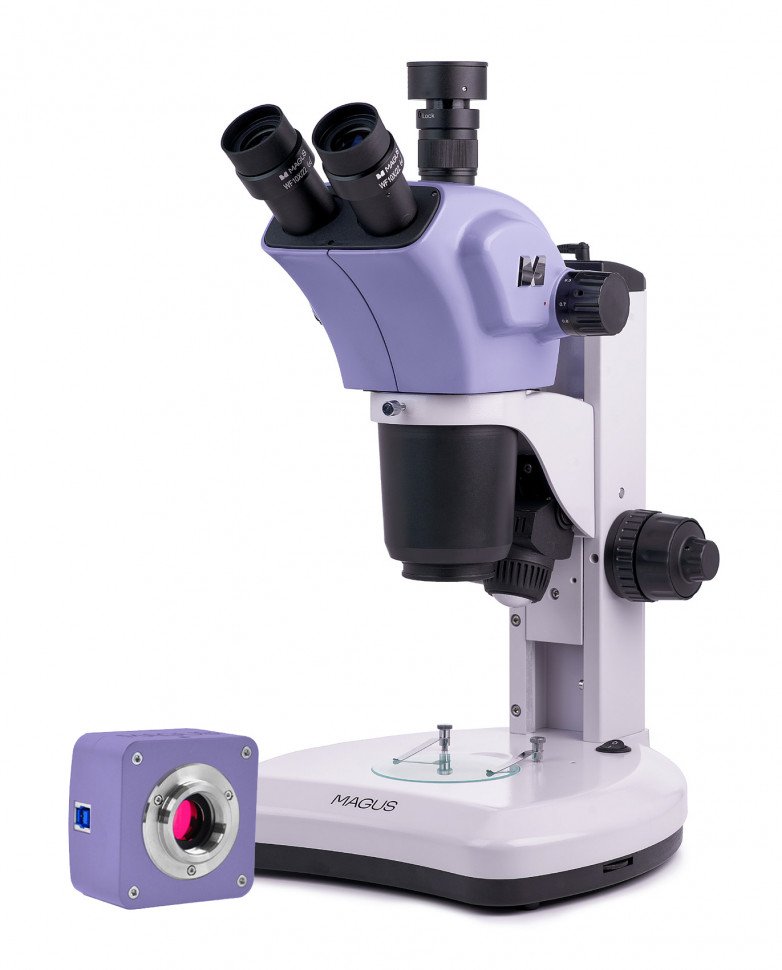
Digital stereoscopic microscope MAGUS Stereo D9T
The MAGUS Stereo D9T stereo microscope is designed for observing three-dimensional objects and details of their structure while maintaining virtual volume and clarity of surface relief. The microscope is equipped with built-in transmitted and reflected light illuminators for examining transparent and opaque objects. Option available for operation in polarized light. The Greenough optical design provides a high depth of field and good three-dimensionality of the image due to the stereoscopic angle of 15 degrees. The microscope magnifies the image of an object without losing spatial orientation; it is comfortable to carry out micromanipulations with objects in a working area of 110–188 mm, so the microscope is used for restoration, soldering, assembly, quality control and other similar operations. Digital camera The MAGUS CBF10 digital camera is designed to work using the bright field method. The camera is equipped with an 18 megapixel sensor and produces a realistic image in a resolution of 4912x3684 pixels. The camera is recommended for use with 4x and 10x lenses of a flat field microscope and with a stereo microscope. When working with low magnification lenses, the camera will allow you to distinguish more fine details. At maximum resolution, video is recorded at a frequency of 13.1 frames per second. This is enough to fine-tune the focus of the microscope. At resolutions of 2456x1842 pixels and 1228x922 pixels, the frequency increases to 34.3 and 54.4 frames per second, respectively. The transitions between frames become softer, the camera records very small movements of the drug. These modes are suitable for demonstrations in the classroom, recording videos of fast-moving processes, and monitoring moving objects. The camera is equipped with a USB 3.0 interface. Data transfer speed is 10 times faster than cameras with USB 2.0 interface. The high-speed camera is recommended for professional work in the laboratory, research or teaching at universities. Visual attachment The trinocular visual head rotates 360°. The digital camera is installed in a vertical tube with an imaging channel. A C-mount adapter for installing the camera is included with the microscope. Zoom The lens smoothly changes magnification by 9x without losing focus. The microscope forms a direct (non-inverted) three-dimensional image. Additional lens attachments change the magnification range, field of view and working distance of the microscope. Focus Coaxial coarse and fine focusing knobs are located on both sides of the tripod. Fine focusing is convenient to use for fine adjustments when working at magnifications above 40x. The rigidity of the coarse focusing mechanism is adjustable. Table inserts Depending on the object of observation, a transparent, white or black and white plate is selected. Transparent and white plates provide uniform illumination of the sample and optimal light dispersion when observing transparent and translucent objects. A black and white plate is selected for studying opaque objects against a contrasting background: the black side is for light objects, the white side is for dark ones. Light sources The microscope is equipped with a LED transmitted light illuminator (5 W) and a reflected light illuminator with oblique illumination (3 W). LED service life is 50,000 hours.
- Microscope type: digital, stereoscopic/instrumental
- Attachment type: trinocular
- Nozzle: 360° rotatable
- Eyepiece tilt angle: 45°
- Magnification, x: 7–63 basic equipment (*optional: 3.5–315); 9:1 zoom ratio
- Diameter of the eyepiece tube, mm: 30
- Eyepieces: 10x/22 mm, remote pupil (*optional: 15x/15 mm, 16x/15 mm, 20x/12 mm, 25x/9 mm)
- Objectives: 0.7–6.3x (*optional: with 0.5x attachment: 0.35–3.15x; with 2x attachment: 1.4–12.6x)
- Working distance, mm: 110 (with nozzle 0.5x: 188; with nozzle 2x: 40.4)
- Interpupillary distance, mm: 53–75
- Linear field of view, mm: 31.43–3.49 (with 0.5x attachment: 62.85–6.98; with 2x attachment: 15.71–1.745), with 10x eyepiece
- Stage, mm: O90 (black and white plate, white plate, transparent plate)
- Diopter adjustment of eyepieces, D: ±5 (on each eyepiece)
- Focusing: coaxial, coarse (83 mm, with a rigidity adjustment mechanism) and fine (0.002 mm)
- Backlight: LED
- Brightness adjustment: yes
- Power source: AC, 220 V / 50 Hz
- Backlight type: reflected light: oblique illuminator – LED 3 W; transmitted light: LED 5W
- Weight, kg: no more than 8, without packaging
- Dimensions, mm: no more than 280x463x335 (WxHxD), without packaging
- Number of megapixels: 18
- Sensor: SONY Exmor CMOS, color, 1/2.3" (6.14x4.61 mm)
- Pixel size, microns: 1.25x1.25
- Sensitivity, volts per lux-second at 550 nm: 0.62 mV/lux/s
- Video recording capability: yes
- Frame rate: 13.1@4912x3684; 34.3@2456x1842; 54.4@1228x922
- Place of use: imaging channel, eyepiece tube instead of eyepiece; C-mount
- Spectral range, nm: 380-650 (with IR filter)
- Shutter Type: ERS (Electronic Rolling Shutter)
- White balance: auto/manual
- Exposure control: auto/manual
- Software, drivers: MAGUS View
- Software features: image size, brightness, exposure time
- Output: USB 3.0, 5 Gb/s
- System requirements: Windows 8/10/11 (32 and 64 bit), Mac OS X, Linux, up to 2.8 GHz Intel Core 2 or higher, at least 2 GB of RAM, USB 3.0 port, CD-ROM, 17" monitor or larger
- Housing: solid aluminum
- Camera power supply: DC 5V from computer USB port
- Operating temperature range, °C: –10... 50
- Possibility of connecting other equipment: simple polarization device
- User level: for professionals, for experienced
- Assembly and setup difficulty level: difficult
- Photo: *.jpg, *.bmp, *.png, *.tif
- Video: *.wmv, *.avi, *.h264 (Win 8 or higher), *h265 (Win 10 or higher)
- Optical design: Greenough stereo microscope
- Purpose: for applied work
- Backlight location: combined
- Research method: bright field
- Digital camera included: yes
- Cover/case/bag included: dustproof cover
- Maximum resolution: 4912x3684 pixels