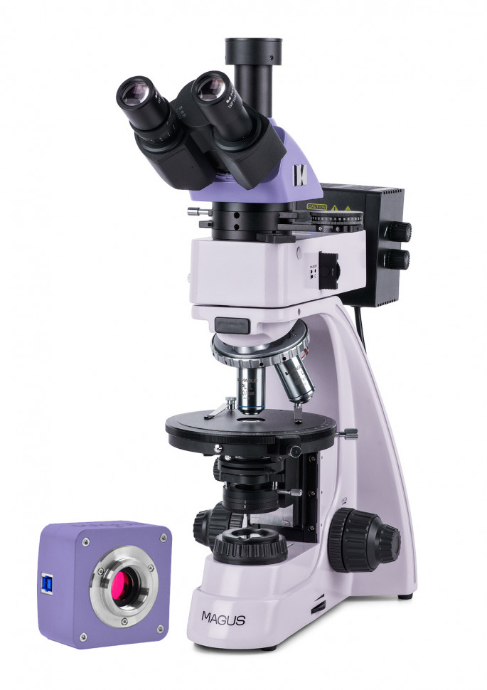
Digital polarizing microscope MAGUS Pol D850
The MAGUS Pol D850 microscope is designed for studying objects in polarized and normal light. Geological sections and thin anisotropic biological and polymer objects are studied in transmitted light. Opaque polished sections with one side polished are examined in reflected light. The thickness of the polished section is arbitrary, usually 5–10 mm. The microscope allows you to study opaque objects up to 15 mm thick. To form an image, a polarizing microscope uses the optical property of birefringence of anisotropic objects. Plane-polarized light, when passing through an anisotropic sample, is split into two beams and changes the plane of polarization. The analyzer brings the oscillations of the beams into one plane, where they interfere. The bright, high-contrast image changes color as the stage rotates. The microscope lenses are free from internal stress. The intermediate tube contains an analyzer and a Bertrand lens, and there is a slot for installing compensators. The microscope is used in crystallography, petrography, mineralogy, forensics, medicine and other fields of science. Digital camera MAGUS CBF70 is a scientific camera with a resolution of 21 megapixels and a 4/3" matrix for working using the bright field method. The camera produces a realistic image in a resolution of 5280x3954 pixels. The camera is recommended for working with 4x, 10x, 20x and 40x lenses. When working with low magnification lenses, the camera will allow you to distinguish more small details. At maximum resolution, video is recorded at a frequency of 17 frames per second. This is enough to fine-tune the focus of the microscope. Reducing the resolution increases the frame rate to 56, 67 and 192 frames per second. The transitions between frames become softer, the camera records very small movements of the drug. These modes are suitable for demonstrations in the classroom, recording videos of fast-moving processes, and monitoring moving objects. The camera is equipped with a USB 3.0 interface. Data transfer speed is 10 times faster than cameras with USB 2.0 interface. The high-speed camera is recommended for professional work in the laboratory, research or teaching at universities. Visual attachment The trinocular visual attachment is designed for infinity. Digital image capture devices are installed in the vertical tube of the imaging channel. The light switch directs 100% of the light to either the digital camera or the eyepiece tubes. Diopter adjustment on the left eyepiece tube. Revolving device Revolver with 5 lenses. The free hole is used to adjust the position of the reflected light illuminator lamp. The laboratory technician installs an additional lens in the free slot of the revolver, which will allow one more magnification. The design of the revolver, tilted toward the tripod (“away from the observer”), frees up space in the front of the stage, and the user sees the lens that introduced the rays. The revolver sockets are centered to align the optical axis of the lens and microscope. Lenses Planachromat infinity objectives are designed specifically for polarized light research: voltage-free optics ensure that birefringence comes from the sample and not from the optical elements. The lenses are designed to operate without a cover glass. Focus Coaxial coarse and fine focusing knobs are located at the bottom of the tripod on either side. The user places his hands freely on the table and takes a relaxed position while working. Focus is adjusted smoothly and effortlessly. The design of the focusing mechanism allows for quick adjustment of the microscope after changing the object of study. To do this, there is a coarse focus locking handle on the right side. By rotating the ring on the left side, you can adjust the comfortable rigidity of the coarse focusing stroke. Subject table The stage rotates 360° to observe the color change of the sample when the polarizer and analyzer are crossed. The rotation angle is marked on the table. Using a vernier, measurements are carried out with an accuracy of 0.1°. The design of the stage provides for centering using two screws, since the analysis of an anisotropic object in polarized light requires exact coincidence of the axis of rotation of the stage with the optical axis of the microscope. Light sources The reflected and transmitted light illuminators contain 30-watt halogen lamps. Halogen lamps emit light at a color temperature that is comfortable for the eyes. The 30-watt lamp is bright enough to work with lenses ranging from 4x to 100x magnification in bright field and polarized light. Indirect Lighting System The lighting system implements the Köhler method. The field and aperture diaphragms are centered in the optical axis at the factory and do not require additional centering. If necessary, the diaphragms are adjusted with centering screws. The light source is centered along three axes. The removable analyzer and polarizer implement the polarization method. The polarizer rotates in the range of 0–360°, the analyzer rotates 360°, and the analyzer has a vernier scale for accurate angle setting. A set of light filters will help you correctly adjust color rendering. Transmitted light lighting system An adjustable field diaphragm, a centered and height-adjustable Abbe condenser with an adjustable aperture diaphragm and a folding Lazo lens realize Köhler illumination adjustment. The numerical aperture of the condenser is 1.25. The polarizer rotates in the range of 0–360°, and the scale has marks for four rotation angles: 0°, 90°, 180°, 270° relative to the analyzer. The analyzer rotates 360°, the analyzer has a vernier scale for precise angle setting. Köhler illumination in reflected and transmitted light Correct adjustment of the microscope according to Köhler improves the quality of the image of the object. With this lighting, the resolution limit on each lens is reached. Uniform illumination of the field of view without darkening at the edges. The object of interest is brought into focus and the artifact images are removed. Observing objects in polarized light The intermediate nozzle contains an analyzer, a Bertrand lens and a slot for installing compensators. The analyzer is introduced into the beam path to observe objects in polarized light. As the polarizer and analyzer rotate, the polarization angle changes. A mutually perpendicular position is achieved by setting both filter fields to the “0” position. When the microscope table is rotated, a change in the refraction of light by the sample is observed depending on the polarization angle. Conoscopic examinations are performed using a Bertrand lens. Compensators are used to enhance the contrast of objects with weak birefringence.
- Microscope type: digital, light/optical, biological
- Attachment type: trinocular
- Nozzle: Siedentopf
- Eyepiece tilt angle: 30°
- Magnification, x: 50–600 basic equipment (*optional: 25–1000/1600/2000)
- Eyepiece tube diameter, mm: 23.2
- Eyepieces: 10x/20 mm; 10x/20 mm with crosshair (*optional: 10x/20 mm with scale; 16x/11 mm; 20x/11 mm)
- Lenses: planachromatic, infinity corrected, stress free: PL 5x/0.12; PL 10x/0.25; PL 40x/0.6; PL 60x/0.7 (*optional: PL 2.5x/0.07; PL 50x/0.7; PL 80x/0.8; PL 100x/0.85); parfocal height 45 mm, designed for working with objects without a cover glass
- Revolver device: for 5 lenses with centered sockets
- Working distance, mm: 26.1 (5x); 5.0 (10x); 3.98 (40x); 2.03 (60x); 11 (2.5x); 3.69 (50x); 1.25 (80x); 0.4 (100x)
- Interpupillary distance, mm: 48–75
- Subject stage, mm: O150, rotated 360°; centered, rotation angle graduated in 1° increments; Vernier for measuring angles with an accuracy of 0.1°
- Diopter adjustment of eyepieces, D: ±5 (on the left tube)
- Condenser: Centerable and height-adjustable Abbe condenser NA 1.25 with adjustable aperture diaphragm and flip-up lens
- Aperture: adjustable aperture, adjustable iris field
- Focusing: coaxial, coarse (21 mm, 39.8 mm/turn, with locking and rigidity adjustment mechanisms) and fine (0.002 mm)
- Backlight: halogen
- Brightness adjustment: yes
- Power source: AC mains, 220±22 V / 50 Hz
- Backlight type: reflected and transmitted light: 12V/30W halogen lamp
- Light filters: reflected light: matte, yellow, green, blue
- Operating temperature range, °C: +5… +35
- Number of megapixels: 21
- Sensor: SONY Exmor CMOS, color, 4/3" (17.4x13.0 mm)
- Pixel size, microns: 3.3x3.3
- Sensitivity, volts per lux-second at 550 nm: 399 mV at 1/30 s
- Video recording capability: yes
- Frame rate: 17@5280x3954; 17@3952x3952; 56@2640x1976; 67@1760x1316; 192@584x438
- Place of use: imaging channel, eyepiece tube instead of eyepiece; C-mount
- Spectral range, nm: 380-650 (with IR filter)
- Shutter Type: ERS (Electronic Rolling Shutter)
- White balance: auto/manual
- Exposure control: auto/manual
- Software, drivers: MAGUS View
- Software features: image size, brightness, exposure time
- Output: USB 3.0, 5 Gb/s
- System requirements: Windows 8/10/11 (32 and 64 bit), Mac OS X, Linux, up to 2.8 GHz Intel Core 2 or higher, at least 2 GB of RAM, USB 3.0 port, CD-ROM, 17" monitor or larger
- Housing: solid aluminum
- Camera power supply: DC 5V from computer USB port
- Operating temperature range, °C: –10... 50
- User level: for professionals, for experienced
- Assembly and setup difficulty level: difficult
- Photo: *.jpg, *.bmp, *.png, *.tif
- Video: *.wmv, *.avi, *.h264 (Win 8 or higher), *h265 (Win 10 or higher)
- Polarizer: transmitted light: with marks 0°, 90°, 180°, 270° on the scale; 360° rotatable; reflected light: removable
- Intermediate attachment: built-in analyzer, switchable to normal or polarized light, rotatable 0–360°; vernier for measuring angles with an accuracy of 0.1°; Bertrand lens; slot for installing compensators
- Compensator: λ compensator; λ/4 compensator; quartz wedge
- Purpose: laboratory/medical
- Backlight location: combined
- Research method: polarization, bright field
- Digital camera included: yes
- Cover/case/bag included: dustproof cover
- Maximum resolution: 5280x3954 pixels