Digital microscope MAGUS Bio DH260
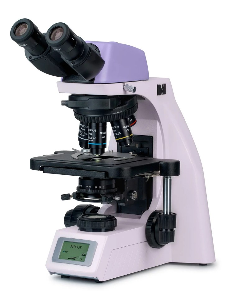
The MAGUS Bio DH260 microscope is equipped with a digital camera: it is built into the visual attachment and allows you not only to make observations through the eyepieces, but also to record an image from the lens or broadcast it on the screen. The MAGUS Bio DH260 is a biological microscope that can be used to examine thin transparent and translucent samples in transmitted light in a bright field. If necessary, the model can be used for research using the dark field method, phase contrast, in polarized or fluorescent light - for this, the microscope must be supplemented with optional parts. Other features of the MAGUS Bio DH260 model are a “smart” lighting system that adjusts the brightness to the selected lens, and an LCD work status screen on which you can clarify the selected study parameters. The microscope is equipped with a visual attachment for 2 eyepieces. The tubes are designed for infinity optics and can change their height - to do this they need to be rotated around their axis. This feature will help the user work more comfortably: the tubes adapt to the user’s height, which means that tension will not accumulate in the neck and shoulder girdle. Diopter adjustment is carried out using complete 10x/22 mm eyepieces. Their exit pupil is placed at a great distance so that the microscope can be used with glasses. Flat rubber eyecups protect the optics of the eyepieces from scratches. The visual attachment has a built-in digital camera. The camera operates on an 8 megapixel matrix and produces materials in 4K resolution (3840x2160 pixels): the drug is visible clearly and distinctly, it can be seen in detail. It is recommended to combine the camera with eyepieces of 4x and 10x magnification. High frame rate – 30 frames per second at maximum resolution – makes video recordings “live”, without gaps between frames. On video, you can effectively track a moving object: not a single detail will escape your gaze. The camera is equipped with a Wi-Fi interface, which can be used to transfer data to a computer. The microscope has a revolving device for 5 objectives with a parfocal height of 60 mm. The microscope kit includes 4 plan achromatic objectives with magnification from 4x to 100x. The user can add the last slot in the revolver at his own discretion. For research in polarized light, an analyzer is installed in the slot of the revolver. A special feature of the revolver is coded lighting. Each lens transmits a different amount of light, which is why when changing magnification, the image in the eyepieces or on the camera is illuminated differently, and the user has to adjust it manually each time. The coded revolver allows you to set the desired lighting for each lens once, and then when you switch lenses, the light will change its intensity automatically. This feature will not only save the user’s time, but also protect his eyes from fatigue due to changes in lighting. Focus is adjusted using coaxial coarse and fine adjustment knobs. Rough adjustments are made with the handle on the left; for fine adjustments, the handles are located on both sides of the tripod. In addition, there is a coarse focus lock knob (used to adjust focus when changing lenses) and a ring that adjusts the coarse focusing stroke. A stage without a retractable rack, but with a belt drive mechanism allows the object to move smoothly across the surface. The user controls the table by manipulating the long handle and placing his hands comfortably on the table. The object under the slide is held in place using a preparation holder, which can be easily removed from the table if necessary. The condenser also has its own characteristics. It can be placed in the desired position vertically and relative to the optical axis. To make it easier to adjust the iris aperture diaphragm, there are marks on the condenser body indicating the magnification of the lenses, and there is an indicator on the ring. To obtain a clear, contrasting image, the pointer must be set to the mark in accordance with the magnification of the working lens. In addition, there is a slot on the condenser body for installing darkfield or phase contrast sliders; when working using other methods, the slot is closed with a plug. The light in the microscope is generated by a three-watt LED with a stable color temperature. Its service life is designed for 50,000 hours. The illuminator allows you to adjust Köhler lighting: it creates a picture that is equally well illuminated throughout the entire field of view, while artifacts do not fall into the field of view. The selected parameters - magnification of the working lens, operating mode, lighting intensity - are displayed on the screen built into the base of the microscope. Parameters are set using two knobs. The design of the microscope is made in such a way as to make work as comfortable as possible for the user. In addition, it is convenient to store or carry: there is a special handle on the body of the device, and the power cord is located so that it is not visible. The microscope can be complemented by a wide range of accessories that expand research capabilities: these are eyepieces and objectives of different magnifications and functionality, components for research in the dark field, phase contrast, fluorescent or polarized light, as well as a calibration slide for measuring objects.
- Microscope type: biological
- Purpose: laboratory
- Magnification, times: 40 - 1000
- Attachment type: binocular
- Microscope attachment: Gemel (Siedentopf with 360° tube rotation)
- Eyepiece tilt angle: 30°
- Diopter adjustment of eyepieces: ±5 diopters on both tubes
- Interpupillary distance, mm: 47 - 78
- Mounting diameter of eyepieces: 30 mm
- Eyepieces: 10x/22 mm
- Lenses: planachromatic, 4x/0.10, 10x/0.25, 40xs/0.65, 100xs/1.25 (oil)
- Lenses/working distance, mm: 30 (4x); 10.2 (10x); 1.5 (40xs); 0.2 (100xs)
- Parfocal height: 60 mm
- Revolving device: for 5 lenses, coded
- Stage: 230x150 mm, two-axis mechanical, without drawer
- Range of movement of the drug, mm: 78 × 54
- Condenser: Centerable and height-adjustable NA 1.25 Abbe condenser with adjustable aperture diaphragm and plug slot for darkfield and phase contrast slider
- Condenser mount: dovetail
- Diaphragm: field, aperture
- Köhler lighting: yes
- Additionally: automatic adjustment of lighting brightness when changing lenses, status display on the screen, sleep mode, eco mode
- Focusing: coaxial, coarse (30 mm, 37.7 mm/rev., with locking and rigidity adjustment mechanisms) and fine (0.002 mm, 0.2 mm/rev.)
- Backlight type: LED
- Backlight type: LED 3W
- Illumination location: bottom
- Backlight power: mains 220 V/50 Hz
- Examination method: bright field, dark field (optional), phase contrast (optional), polarization (optional)
- Optional modules: dark field slider, simple polarization device (polarizer and analyzer), phase contrast device (slider and lenses)
- Number of megapixels: 8
- Sensor type: CMOS, color
- Matrix size: 1/1.8''
- Pixel size, microns: 2
- Frame rate, 1/s: 30
- Maximum resolution, pixel: 3840 × 2160
- Country of origin: China
- Manufacturer's warranty: 5 years
- Package size: 47 × 32 × 67 cm
- Package weight: 12.1 kg