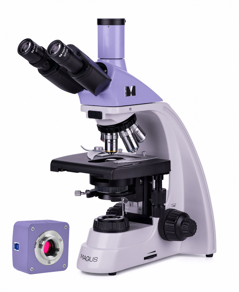
Digital microscope MAGUS Bio D230T
The MAGUS Bio D230T microscope is designed for observing transparent and translucent biological samples in the form of smears and sections in transmitted light in a bright field. Installation of optional components will allow the use of dark field, phase contrast and polarization methods. The microscope is suitable for routine laboratory work, scientific research, and teaching. Digital camera The MAGUS CDF10 digital camera is designed to work using light and dark field methods. It is characterized by low noise level and high light sensitivity. The camera is equipped with a 2 megapixel sensor and produces a realistic image in Full HD resolution (1920x1080 pixels) when working with 40x, 60x and 100x lenses. Video recording is carried out at a frequency of 125 frames per second at maximum resolution. The videos are smooth, with soft and invisible transitions between frames. The movement of the drug is displayed in real time, without delays. The camera provides convenient work with moving objects and is suitable for conducting demonstrations in the classroom. The camera is equipped with a USB 3.0 interface. Data transfer speed is 10 times faster than cameras with USB 2.0 interface. The high-speed camera is recommended for professional work in the laboratory, research or teaching at universities. Visual attachment Trinocular imaging head with infinity-rated optics. The nozzle tubes rotate 360°. The user selects the eye relief height in accordance with his own height. The digital camera is installed in a vertical tube with an imaging channel. Revolving device Revolver with 5 lenses. A free revolver socket for installing an additional lens, which will allow you to get another magnification within the magnification range. The revolver is turned “away from the observer”: the user sees the lens that introduced the rays, the space above the table is free. Focus Coaxial coarse and fine focusing knobs are located at the bottom of the body on both sides of the tripod. The user places his hands freely on the table and takes a relaxed position while working. Focus adjustment is smooth and effortless. Subject table The table does not have a retractable rack along the X axis, which increases the ergonomics of operation. The belt drive mechanism moves the object smoothly. The drug holder is secured with two screws and can be removed during quick manual scanning. Abbe condenser The condenser has a slot for installing a dark field slider or a phase contrast slider. Installing a slider saves time when switching from one research method to another. Light source The halogen lamp emits warm tones that are familiar to the eye. Replacing the lamp requires minimal effort. The 30-watt lamp produces enough brightness to work with all brightfield, darkfield and phase contrast lenses. Accessories A line of accessories has been developed for the microscope: eyepieces, lenses, devices for working using the dark field method, phase contrast and polarization, digital cameras, calibration slides.
- Microscope type: digital, light/optical, biological
- Attachment type: trinocular
- Nozzle: Gemel (Siedentopf with 360° rotation of tubes)
- Eyepiece tilt angle: 30°
- Magnification, x: 40–1000 basic equipment (*optional: 40–1500/1600/2000)
- Eyepiece tube diameter, mm: 23.2
- Eyepieces: 10x/18, eye relief 10 mm (*optional: 15x/11 mm, 16x/11 mm, 20x/11 mm)
- Lenses: achromatic, infinity corrected: 4x/0.1; 10x/0.25; 40xs/0.65; 100xs/1.25 (oil); parfocal height 45 mm (*optional: 20x/0.4)
- Revolving device: for 5 lenses
- Working distance, mm: 18.89 (4x); 5.95 (10x); 0.775 (40xs); 0.36 (100xs); 2.61 (20x)
- Interpupillary distance, mm: 48–75
- Subject stage, mm: 180x150, two-axis mechanical, without retractable rail
- Range of movement of the object table, mm: 75/50
- Diopter adjustment of eyepieces, D: ±5 (on the left tube)
- Condenser: Centerable and height-adjustable NA 1.25 Abbe condenser with adjustable aperture diaphragm and slot for darkfield and phase contrast slider; dovetail fastening type
- Aperture: adjustable aperture, adjustable iris field
- Focusing: coaxial, coarse (21 mm, 39.8 mm/turn, with locking and rigidity adjustment mechanisms) and fine (0.002 mm)
- Backlight: halogen
- Brightness adjustment: yes
- Power source: AC mains, 220±22 V / 50 Hz
- Backlight type: 12 V/30 W, G4 base
- Light filters: yes
- Weight, kg: no more than 8, without packaging
- Dimensions, mm: no more than 200x436x400 (WxHxD), without packaging
- Operating temperature range, °C: +5… +35
- Number of megapixels: 2
- Sensor: SONY Exmor CMOS, color, 1/2" (7.2x4.05 mm)
- Pixel size, microns: 3.75x3.75
- Sensitivity, volts per lux-second at 550 nm: 2350 mV at 1/30 s
- Video recording capability: yes
- Frame rate: 125@1920x1080
- Place of use: imaging channel, eyepiece tube instead of eyepiece; C-mount
- Spectral range, nm: 380–650 (with IR filter)
- Shutter Type: ERS (Electronic Rolling Shutter)
- White balance: auto/manual
- Exposure control: auto/manual
- Software, drivers: MAGUS View
- Software features: image size, brightness, exposure time
- Output: USB 3.0, 5 Gb/s
- System requirements: Windows 8/10/11 (32 and 64 bit), Mac OS X, Linux, up to 2.8 GHz Intel Core 2 or higher, at least 2 GB of RAM, USB 3.0 port, CD-ROM, 17" monitor or larger
- Housing: solid aluminum
- Camera power supply: DC 5V from computer USB port
- Operating temperature range, °C: –10... 50
- Possibility of connecting other equipment: phase contrast device (condenser and objectives), dark field condenser (dry or oil), simple polarization device (polarizer and analyzer)
- User level: for professionals, for experienced
- Assembly and setup difficulty level: difficult
- Photo: *.jpg, *.bmp, *.png, *.tif
- Video: *.wmv, *.avi, *.h264 (Win 8 or higher), *h265 (Win 10 or higher)
- Purpose: laboratory/medical
- Illumination location: bottom
- Research method: bright field
- Digital camera included: yes
- Cover/case/bag included: dustproof cover
- Maximum resolution: 1920x1080 pixels