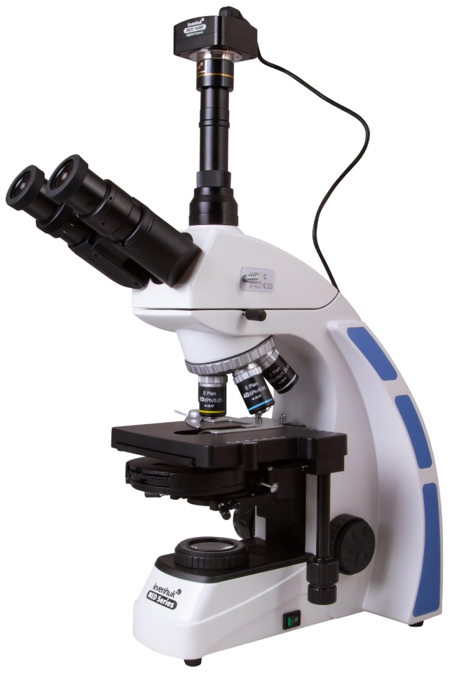
Levenhuk MED D45T Digital Microscope
Levenhuk MED D45T digital microscope is equipped with a 16 MP digital camera, which allows you to simultaneously monitor and broadcast the image to a PC. Using the phase contrast method increases the saturation and clarity of transparent objects without additional coloring. This feature is extremely useful when studying microbiological living samples that will die when paint is applied. This device is an excellent purchase for both a scientific laboratory and a medical institution. What are the advantages of a digital model? By connecting the camera to a PC using a USB cable, you can broadcast the research process online to an external monitor. Thus, the Levenhuk MED D45T digital microscope can become a visual aid in the educational process, and also allows you to involve a group of people in research. For further processing of images and videos, the package includes a CD with software that allows you to change the color parameters of the image. In addition, using a wide screen rather than a lens as a means of observation, you will reduce eye strain and be able to see the smallest details. High-quality optics with infinite correction type As a professional category device, a digital microscope is equipped with an optical system, the infinite type of correction of which allows additional auxiliary elements, for example, illuminators or polarizers, to be built into the optical space between the tube and the lenses. Thus, the Levenhuk MED D45T biological microscope can be optimized to solve a wide range of research problems. Thoughtful design and convenient operation Thanks to the possibility of tilting the viewing part by 30°, you will not have to strain your neck and shoulders during long-term studies. All optical elements of the device have a coating that protects against fungus, which is very useful for biological research. For convenient and prompt placement of the material under study under the lens, the object stage of the device is equipped with a preparation guide. You can also install additional filters in a special holder to change the contrast and improve detail.
- Microscope type: biological
- Attachment type: trinocular
- Maximum resolution: 4632x3488
- Optics material: optical glass with anti-fungal coating
- Nozzle: 360° rotatable
- Eyepiece tilt angle: 30°
- Magnification, times: 40-1000
- Diameter of the eyepiece tube, mm: 30 mm (binocular attachment); 23.2 mm (third vertical tube)
- Eyepieces: wide-field with diopter adjustment WF 10x/22 mm (2 pcs.)
- Lenses: phase plan achromatic, infinity corrected: 4x, 10x, 40xs, 100xs (oil)
- Revolving device: for 5 lenses
- Interpupillary distance, mm: 48–75
- Subject table, mm: mechanical two-layer, 180x150 mm, with preparation guide
- Range of movement of the object table, mm: 75/50
- Diopter adjustment of eyepieces, D: ±5
- Condenser: phase (dark field)
- Focusing: coaxial, coarse (0.5 mm) and fine (0.002 mm), with rack and pinion mechanism
- Case: metal
- Backlight: LED
- Brightness adjustment: yes
- Backlight type: 5W AC 85-230V
- Light filters: blue, green, yellow
- Additionally: collector, Koehler lighting
- Number of megapixels: 16
- Sensing element: 1/2.33
- Pixel size, microns: 1.335x1.335
- Video recording capability: yes
- Frame rate: 2@4632x3488 8@2320x1740 11@1536x1160
- Use location: 23.2mm microscope third eyepiece tube
- Spectral range, nm: 380–650
- White balance: auto/manual
- Exposure control: 0.2–2000 ms
- Software, drivers: Levenhuk
- Software features: image size, brightness, exposure time
- Output: USB 2.0, 480 Mbps
- System requirements: Windows XP/Vista/7/8/10 (32 and 64 bit), Mac OS 10.12, Linux, 2.8 GHz Intel Core 2 or higher, at least 2 GB RAM, USB 2.0, 17 monitor
- Camera power supply: via USB cable
- Purpose: laboratory/medical
- Illumination location: bottom
- Research method: phase contrast microscopy, dark field, bright field
- Digital camera included: yes
- Power supply: 100-240V