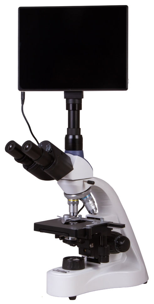
Levenhuk MED D10T LCD Digital Microscope
Levenhuk MED D10T LCD is a digital microscope that is interesting not only for its optical capabilities, but also for its camera with a 5-megapixel sensor and LCD screen. The microscope is suitable for visual observations and for creating digital archives of observations. The capabilities of this optical device will be useful in a medical laboratory, research center, or university department. A microscope (LCD) is worth buying if you are involved in professional research and spend many hours studying microspecimens. The LCD screen significantly reduces eye strain. High-end achromatic optics The trinocular attachment is divided into a binocular visual part and a separate vertical tube into which the digital camera is installed. The nozzle can be rotated 360°, and the visual unit is tilted at a 30-degree angle. For observations, wide-field eyepieces with diopter correction are used, which provide 10x magnification. The revolving device houses four achromatic lenses that convey a detailed and contrasting image. The 40x and 100x lenses have spring-loaded frames; the 100x lens is used for immersion observations. The optical surfaces of the eyepieces and lenses are protected with an anti-fungal coating. Digital capabilities of the microscope A digital camera with a 5-megapixel sensitive sensor is designed for photo and video shooting. Highly detailed images and smooth video speed allow them to be used for professional research. The image from the microscope lens is displayed on the built-in LCD screen, so no additional equipment is required to use the camera. The camera can work with different accessories: keyboard, headphones, external monitor, memory cards. A special program makes it possible to edit the footage. Easy and convenient work in the laboratory The camera makes work in the laboratory easier and more convenient for the researcher - there is no need to constantly strain your eyes while looking at samples through eyepieces: everything can be seen on the built-in LCD screen with touch control. Using the software, you can adjust the size, brightness, contrast, gamma, sharpness and saturation of the image, change the exposure time and white balance, calibrate the camera with lenses, measure samples or individual structures (several units of measurement are available). In addition, the program allows for particle analysis. Bright illumination of samples with a powerful LED Microspecimens are mounted on a two-coordinate stage, under which there is a bright LED backlight. Also located underneath is an Abbe condenser with an iris diaphragm and a filter holder. There are coarse and fine focusing knobs nearby. The brightness of the backlight is adjustable. An AC power connection is required to power the backlight.
- Microscope type: biological, digital
- Purpose: laboratory, medical
- Maximum resolution, pixel: 2048×1536
- Number of megapixels: 5
- Sensor type: CMOS
- Matrix size: 1/2.5"
- Pixel size, microns: 2.2
- Sensitivity at λ= 550 nm, V/lx-s: 0.53
- Magnification, times: 40 - 1000
- Optics material: optical glass with anti-fungal coating
- Attachment type: trinocular
- Rotation of the eyepiece: yes, 360°
- Eyepiece tilt angle: 30°
- Diopter adjustment of eyepieces: ±5 diopters
- Interpupillary distance, mm: 48 - 75
- Mounting diameter of eyepieces: 30 mm
- Display: 9.4 inches, color, touch
- Additionally: internal diameter vert. tube: 23.2 mm
- Eyepieces: wide field 10x/18 mm
- Lenses: achromatic, 4x/0.10, 10x/0.25, 40xs/0.65, 100xs/1.25 (oil)
- Correction for microscope tube length: 160 mm
- Correction for cover glass thickness: 0.17 mm
- Revolving device: for 4 lenses
- Subject table: 125x130 mm, mechanical two-axis, with specimen guide and coaxially located control handles
- Range of movement of the drug, mm: 70 × 50
- Condenser: Abbe, centered, height adjustable, with filter holder, largest numerical aperture: 1.25
- Aperture: aperture
- Focusing: fine, coarse, coaxially located control knobs, with a height limiter for the table movement
- Fine focusing step, mm: 0.002
- Case material: metal
- Backlight type: LED
- LED power: 5W
- Illumination location: bottom
- Brightness adjustment: yes
- Backlight power: mains 220 V/50 Hz
- Research method: bright field
- Light filters included: blue, green, yellow
- Digital camera included: yes
- Place of use: 23.2mm microscope third eyepiece tube; using a camera mount
- White balance: auto/manual
- Exposure control: auto/manual
- Frame rate: 15 fps
- Software, drivers: Android 5.1 (multilingual)
- Software capabilities: measurements, brightness, particle analysis, etc.
- Possibility of connecting additional equipment: microSD memory cards up to 32 GB; monitor/TV with HDMI connector; flash drive, computer mouse, keyboard with USB connector; 3.5 mm headphones
- Image format: *.jpg
- Video format: 1080p, *.3gp
- Output: USB 2.0 (2 pcs.), miniHDMI, Wi-Fi, TF memory card slot
- Camera power supply: DC 12V; via AC adapter
- Built-in memory: 4 GB
- Country of origin: China
- Manufacturer's Warranty: Lifetime
- Package weight: 6.66 kg
- Package size: 57 × 38 × 26 cm