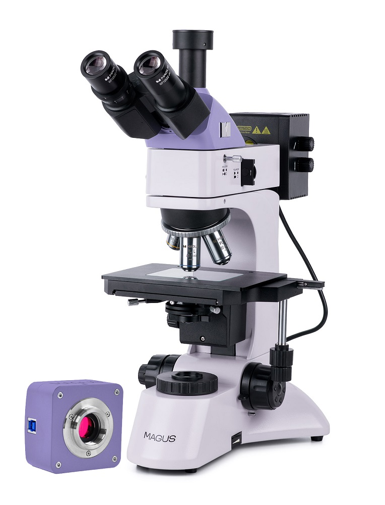
Digital metallographic microscope MAGUS Metal D600
The MAGUS Metal D600 microscope is designed for studying the microstructure of metals, alloys, semiconductor materials, paint coatings and other opaque objects on plane-parallel polished sections. The reflected light illuminator operates using the bright field method and simple polarization. The built-in transmitted light illuminator is used for studying translucent films and objects on filters: samples of air, water, oil, etc. The microscope is used at enterprises of the metallurgical, mechanical engineering, aerospace, nuclear and energy industries, in research laboratories and technical universities. Digital camera The MAGUS CBF30 digital camera is designed to work using the bright field method. The camera is equipped with a 6.3 megapixel sensor and produces a realistic image in a resolution of 3072 x 2048 pixels. The camera is recommended for use with 4x, 10x, 20x and 40x lenses. When working with low magnification lenses, the camera will allow you to distinguish more fine details. Video recording is carried out at a frequency of 59 frames per second at maximum resolution. The videos are smooth, with soft and invisible transitions between frames. The movement of the drug is displayed in real time, without delays. The camera provides convenient work with moving objects and is suitable for conducting demonstrations in the classroom. If necessary, the resolution can be reduced to 1536 x 1024 pixels. The camera is equipped with a USB 3.0 interface. Data transfer speed is 10 times faster than cameras with USB 2.0 interface. The high-speed camera is recommended for professional work in the laboratory, research or teaching at universities. Visual attachment The trinocular visual attachment is designed for infinity. The digital camera is installed in a vertical tube with an imaging channel. The light output switch allows you to direct 100% of the light to either the digital camera or the eyepiece tubes. Diopter adjustment on the left eyepiece tube. Revolving device Revolver with 5 lenses. The free hole is used to adjust the position of the reflected light illuminator lamp. You can also install an additional lens in the free slot of the revolver, which will allow you to get another magnification. The revolver is turned “away from the observer”: the space above the table is free and the user sees the lens that introduced the rays into the path. Lenses Planachromat lenses with an extended working distance are designed for bright field work. Focus Coaxial coarse and fine focusing knobs are located at the bottom of the body on both sides of the tripod. The user places his hands freely on the table and takes a relaxed position while working. Focus adjustment is smooth and effortless. On the right side there is a coarse focusing locking handle, on the left there is a ring for adjusting the rigidity of the coarse focusing stroke. Subject table The object is moved by moving the table surface along two axes. The maximum height of the observed object is 20 mm. The glass insert in the table is used for working with translucent samples. Light sources The reflected and transmitted light illuminators contain 30-watt halogen lamps. Halogen lamps emit light at a color temperature that is comfortable for the eyes. The 30-watt lamp is bright enough to work with lenses ranging from 4x to 100x magnification in bright field and polarized light. Indirect Lighting System The microscope is equipped with a built-in analyzer and a removable polarizer. The polarizer rotates in the range 0° - 360°, the analyzer does not rotate. Using the aperture diaphragm and field diaphragm, lighting is adjusted using the Köhler method. Both apertures and the light source are centered. There is a set of light filters. Transmitted light lighting system An adjustable field diaphragm and a centered and height-adjustable Abbe condenser with a numerical aperture of 1.25 provide Köhler illumination. The condenser has a folding lens for working with low magnification lenses. Accessories A line of accessories has been developed for the microscope: eyepieces, lenses, digital cameras, calibration slides.
- Microscope type: digital, light/optical, metallographic
- Attachment type: trinocular
- Nozzle: Siedentopf
- Eyepiece tilt angle: 30°
- Magnification, x: 50–600 basic equipment (*optional: 50–1000/1250/1500/2000/2500)
- Diameter of the eyepiece tube, mm: 30
- Eyepieces: 10x/22, distance pupil 10 mm (*optional: 10x/22 mm with scale, 12.5x/14 mm, 15x/15 mm, 20x/12 mm, 25x/9 mm)
- Lenses: plan achromatic, infinity corrected: PL L5x/0.12, PL L10x/0.25, PL L40x/0.60, PL L60x/0.70; parfocal height 45 mm (*optional: PL L20x/0.40, PL L50x/0.70, PL L80x/0.80, PL L100x/0.85 (dry))
- Revolving device: for 5 lenses
- Working distance, mm: 26.1 (5x); 20.2 (10x); 3.98 (40x); 2.08 (60x); 8.80 (20x); 3.68 (50x); 1.25 (80x); 0.40 (100x)
- Interpupillary distance, mm: 48–75
- Subject stage, mm: 210x140, two-axis mechanical, with rectangular glass insert
- Range of movement of the object table, mm: 75/50
- Diopter adjustment of eyepieces, D: ±5 (on the left tube)
- Condenser: transmitted light: centered and height-adjustable Abbe condenser NA 1.25 with adjustable aperture diaphragm and flip-up lens; screw fastening type
- Aperture: reflected and transmitted light: built-in aperture and field
- Focusing: coaxial, coarse (25 mm, with locking and rigidity adjustment mechanisms) and fine (0.002 mm)
- Backlight: halogen
- Brightness adjustment: yes
- Power source: AC mains, 220±22 V / 50 Hz
- Backlight type: reflected and transmitted light: 12V/30W
- Light filters: reflected light: matte, yellow, green, blue
- Weight, kg: no more than 11, without packaging
- Operating temperature range, °C: +5… +35
- Additionally: reflected light: built-in analyzer and removable polarizer
- Number of megapixels: 6.3
- Sensor: SONY Exmor CMOS, color, 1/1.8" (7.37x4.92 mm)
- Pixel size, microns: 2.4x2.4
- Sensitivity, volts per lux-second at 550 nm: 425 mV at 1/30 s
- Video recording capability: yes
- Frame rate: 59@3072x2048; 59@1536x1024
- Place of use: imaging channel, eyepiece tube instead of eyepiece; C-mount
- Spectral range, nm: 380-650 (with IR filter)
- Shutter Type: ERS (Electronic Rolling Shutter)
- White balance: auto/manual
- Exposure control: auto/manual
- Software, drivers: MAGUS View
- Software features: image size, brightness, exposure time
- Output: USB 3.0, 5 Gb/s
- System requirements: Windows 8/10/11 (32 and 64 bit), Mac OS X, Linux, up to 2.8 GHz Intel Core 2 or higher, at least 2 GB of RAM, USB 3.0 port, CD-ROM, 17" monitor or larger
- Housing: solid aluminum
- Camera power supply: DC 5V from computer USB port
- Operating temperature range, °C: –10... 50
- User level: for professionals, for experienced
- Assembly and setup difficulty level: difficult
- Photo: *.jpg, *.bmp, *.png, *.tif
- Video: *.wmv, *.avi, *.h264 (Win 8 or higher), *h265 (Win 10 or higher)
- Purpose: metallographic
- Backlight location: combined
- Research method: polarization, bright field
- Digital camera included: yes
- Cover/case/bag included: dustproof cover
- Maximum resolution: 3072x2048 pixels