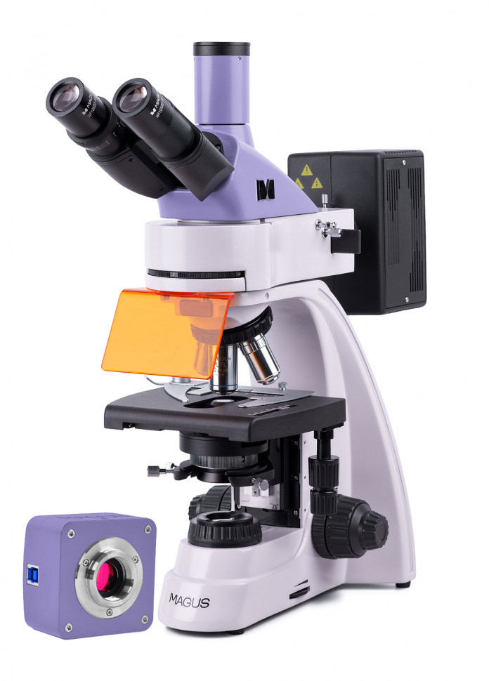
Digital fluorescence microscope MAGUS Lum D400
The MAGUS Lum D400 microscope is designed to study objects when illuminated with reflected light using the luminescence method and when illuminated with transmitted light using the bright field method, and with additional equipment – using dark field, simple polarization and phase contrast methods. The principle of the luminescence method is based on the ability of certain substances to glow when exposed to light from a certain part of the spectrum. Objects are irradiated with invisible short-wave ultraviolet light or violet, blue and green light. The wavelength of the glow is greater than the wavelength of the exciting light. The object glows blue, cyan, green-yellow or red, respectively. Some objects glow on their own, some - after treatment with fluorochromes. The causative agents of tuberculosis, chlamydia, rabies, herpes and other diseases are studied using a fluorescent microscope. The microscope is used for DNA analysis and chromosomal analysis, bone marrow and blood smear examinations, forensic and pharmacological examinations, veterinary control studies and sanitary and epidemiological surveillance. Digital camera The MAGUS CLM10 digital camera is designed to work in luminescent light and dark field. The camera is equipped with a 2.3 megapixel sensor and produces a realistic image in a resolution of 1920x1200 pixels. The camera is recommended for working with 40x, 60x and 100x lenses. When working with medium magnification lenses, the camera will allow you to distinguish more fine details. Video recording is carried out at a frequency of 120 frames per second at maximum resolution. The videos are smooth, with soft and invisible transitions between frames. The movement of the drug is displayed in real time, without delays. The camera provides convenient work with moving objects and is suitable for conducting demonstrations in the classroom. The camera is equipped with a USB 3.0 interface. Data transfer speed is 10 times faster than cameras with USB 2.0 interface. The high-speed camera is recommended for professional work in the laboratory, research or teaching at universities. Visual attachment Trinocular imaging head with infinity-rated optics. The nozzle tubes rotate 360°. The user selects the eye relief height in accordance with his own height. The digital camera is installed in a vertical tube with an imaging channel. Revolving device Revolver with 5 lenses. The free hole is used to adjust the position of the reflected light illuminator lamp. You can also install an additional lens in the free slot of the revolver, which will allow you to get another magnification. The revolver is turned “away from the observer”: the space above the table is free and the user sees the lens that introduced the rays into the path. Lenses Planachromat and microfluar infinity objectives. The standard equipment includes 4x, 10x and 100x lenses, which are used for working in the bright field method, and a 40x lens for working in visible luminescence light. Focus Coaxial coarse and fine focusing knobs are located at the bottom of the body on both sides of the tripod. The user places his hands freely on the table and takes a relaxed position while working. Focus is adjusted smoothly and effortlessly. On the right side there is a coarse focus locking handle for quickly adjusting the microscope after changing the object of study. On the left is a ring for adjusting the rigidity of the coarse focusing stroke for comfortable individual settings. Subject table The table does not have a retractable rack along the X axis, which increases the ergonomics of operation. The belt drive mechanism moves the object smoothly. The drug holder is secured with two screws and can be removed during quick manual scanning. When working in reflected light, a black insert is installed in the table to remove stray light from transmitted light. Luminescent nozzle The luminescence excitation source is a 100-watt mercury lamp. It has high brightness and a wide range of wavelengths. The spectrum of the lamp has peaks, which allows you to work with a variety of fluorescent dyes. The attachment contains four excitation filters: ultraviolet (UV), violet (V), blue (B), green (G). The mercury lamp is located in the flashlight and is centered in three planes. The flashlight removes heat radiation, the lamp does not overheat. Convenient lamp mounting ensures safe and quick replacement. Köhler illumination in reflected and transmitted light Correct adjustment of the microscope according to Köhler improves the quality of the image of the object. With this lighting, the resolution limit on each lens is reached. Uniform illumination of the field of view without darkening at the edges. The object of interest is brought into focus and the artifact images are removed. Condenser The condenser has a slot for installing a darkfield or phase contrast slider. Installing a slider saves time when switching research methods in transmitted light. Transmitted light source The transmitted light illuminator contains a 30-watt halogen lamp. The halogen lamp emits light at a color temperature that is comfortable for the eyes. The 30-watt lamp shines bright enough to work on lenses with magnifications from 4x to 100x.
- Microscope type: digital, light/optical, biological
- Attachment type: trinocular
- Nozzle: Gemel (Siedentopf with 360° rotation of tubes)
- Eyepiece tilt angle: 30°
- Magnification, x: 40–1000 basic equipment (*optional: 40–1250/1500/2000/2500)
- Diameter of the eyepiece tube, mm: 30
- Eyepieces: 10x/22 mm, distance pupil 10 mm (*optional: 10x/22 mm with scale; 12.5x/14 mm, 15x/15 mm, 20x/12 mm, 25x/9 mm)
- Objectives: planachromatic and microfluor, infinity corrected: PL 4x/0.10, PL 10x/0.25, PL FL 40x/0.85, PL 100x/1.25 (mi); parfocal height 45 mm (*optional: PL FL 10x/0.35, PL 60x/0.80?/0.17)
- Revolving device: for 5 lenses
- Working distance, mm: 19.8 (4x); 5.0 (10x); 0.42 (FL 40x); 0.36 (100x); 2.37 (FL 10x); 0.46 (60x)
- Interpupillary distance, mm: 48–75
- Subject stage, mm: 180x150, two-axis mechanical, without retractable rail
- Range of movement of the object table, mm: 75/50
- Condenser: Centerable and height-adjustable NA 1.25 Abbe condenser with adjustable aperture diaphragm and slot for darkfield and phase contrast slider; dovetail fastening type
- Aperture: adjustable aperture, adjustable iris field
- Focusing: coaxial, coarse (21 mm, 39.8 mm/turn, with locking and rigidity adjustment mechanisms) and fine (0.002 mm)
- Backlight: fluorescent, halogen
- Brightness adjustment: yes
- Power Source: AC 85-265V/50/60Hz
- Backlight type: reflected light: mercury lamp 100 W; transmitted light: halogen lamp 12 V/30 W
- Operating temperature range, °C: +5… +35
- Number of megapixels: 2.3
- Sensor: SONY Exmor CMOS, monochrome, 1/1.2" (11.25x7.03 mm)
- Pixel size, microns: 5.86x5.86
- Sensitivity, volts per lux-second at 550 nm: 1016 mV at 1/30 s
- Video recording capability: yes
- Frame rate: 120@1920x1200
- Place of use: imaging channel, eyepiece tube instead of eyepiece; C-mount
- Spectral range, nm: 380–650 (with IR filter and anti-glare filter)
- Shutter type: Global shutter
- White balance: auto/manual
- Exposure control: auto/manual
- Software, drivers: MAGUS View
- Software features: image size, brightness, exposure time
- Output: USB 3.0, 5 Gb/s
- System requirements: Windows 8/10/11 (32 and 64 bit), Mac OS X, Linux, up to 2.8 GHz Intel Core 2 or higher, at least 2 GB of RAM, USB 3.0 port, CD-ROM, 17" monitor or larger
- Housing: solid aluminum
- Camera power supply: DC 5V from computer USB port
- Operating temperature range, °C: –10... 50
- Possibility of connecting other equipment: phase contrast device (condenser and objectives), dark field condenser (dry or oil), simple polarization device (polarizer and analyzer), dark field slider
- User level: for professionals, for experienced
- Assembly and setup difficulty level: difficult
- Photo: *.jpg, *.bmp, *.png, *.tif
- Video: *.wmv, *.avi, *.h264 (Win 8 or higher), *h265 (Win 10 or higher)
- Fluorescent module: filters: ultraviolet (UV), violet (V), blue (B), green (G)
- Purpose: laboratory/medical
- Backlight location: combined
- Research method: fluorescence, bright field
- Digital camera included: yes
- Cover/case/bag included: dustproof cover
- Maximum resolution: 1920x1200 pixels