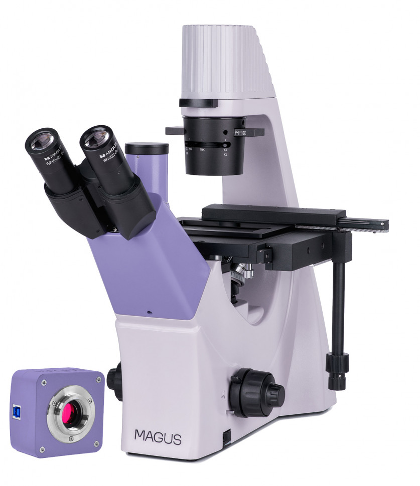
Digital inverted microscope MAGUS Bio VD300
The MAGUS Bio VD300 microscope is designed for the study of liquid sediments, cell colonies, living cells, tissue cultures and other stained and unstained objects in laboratory glassware. Observations are carried out in transmitted light, research methods are bright field and phase contrast. The inverted design of the microscope involves the use of Petri dishes, multi-well plates, vials, roller bottles and flasks up to 70 mm high with a bottom thickness of 1.2 mm. The microscope uses special lenses to work with such glassware. The microscope is used for teaching in universities, routine laboratory work and scientific research. Fields of science: medicine, biotechnology, biology, virology, agriculture, ecology, toxicology, pharmacology and others. Digital camera The MAGUS CDF30 digital camera is designed to work using light and dark field methods. It is characterized by low noise level and high light sensitivity. The camera is equipped with an 8.3 megapixel sensor and produces a realistic image in 4K resolution (3840x2160 pixels), in which very small details of objects and their structures are clearly visible. The camera is recommended for use with 4x, 10x and 20x lenses, as well as a stereo microscope. At maximum resolution, video is recorded at a frequency of 45 frames per second. This is enough for conducting demonstrations in the classroom, fine-tuning the focus of a microscope, recording fast processes and observing moving objects. At Full HD resolution (1920x1080 pixels), the frame rate increases to 70 frames per second. The transitions between frames become softer, the camera records very small movements of the drug. The camera is equipped with a USB 3.0 interface. Data transfer speed is 10 times faster than cameras with USB 2.0 interface. The high-speed camera is recommended for professional work in the laboratory, research or teaching at universities. Visual attachment The optics of the trinocular visual attachment are designed for infinity. The digital camera is installed in a vertical tube with an imaging channel. Light division 50/50. Revolving device The revolver for 4 lenses is mounted on a tripod under the table. Lenses Planachromatic infinity lenses with an increased working distance are adjusted to the thickness of the pan bottom of 1.2 mm. 3 lenses are used to work using the bright field method and one lens is used to work using the phase contrast method. Focus Coaxial coarse and fine focusing knobs are located at the bottom of the body on both sides of the tripod. The user places his hands freely on the table and takes a relaxed position while working. Focus is adjusted smoothly and effortlessly. On the right side there is a coarse focus locking handle for quickly adjusting the microscope after changing the object of study. On the left is a ring for adjusting the rigidity of the coarse focusing stroke for comfortable individual settings. Subject table A special mechanism for moving laboratory glassware in two mutually perpendicular directions is installed on a fixed object stage. Smooth, subtle movement of the object will ensure the accuracy of the research - not a single section of the object under study will be missed. The travel handle is low, so the user does not strain his right hand. The microscope kit includes three cup holders for different sizes of dishes. Condenser A phase contrast slider is installed in the condenser. The phase rings are centered. Using a slider saves time when switching from one research method to another. Light source The 9W LED provides bright subject illumination, sufficient for brightfield and phase contrast work on all lenses. Color temperature does not change when brightness changes. LED service life is 50,000 hours. Accessories A line of accessories has been developed for the microscope. Additional 20x and 40x phase objectives are used for studying non-contrast objects with sizes of 0.7 µm and 0.4 µm. Eyepieces extend the magnification range of the microscope. An additional pair of eyepieces will help realize useful magnification on a lens that is used more often for work.
- Microscope type: digital, light/optical, biological
- Attachment type: trinocular
- Nozzle: Siedentopf
- Eyepiece tilt angle: 45°
- Magnification, x: 100–400 basic equipment (*optional: 100–500/600/800/1000)
- Diameter of the eyepiece tube, mm: 30
- Eyepieces: 10x/22 mm, distance pupil 10 mm (*optional: 10x/22 mm with scale, 12.5x/14 mm, 15x/15 mm, 20x/12 mm, 25x/9 mm)
- Lenses: plan achromatic, infinity corrected: PL 10x/0.25, PL 20x/0.40, PL 40x/0.60, PHP2 10x/0.25 phase; parfocal height 45 mm (*option: PHP2 20x/0.40 phase, PHP2 40x/0.60 phase)
- Revolving device: for 4 lenses
- Working distance, mm: 4.27 (10x); 8.0 (20x); 3.5 (40x)
- Interpupillary distance, mm: 48–75
- Subject stage, mm: fixed, with a mechanical device for moving the sample; cup holders: 29x77 mm, O90 mm; 34x77.5mm, O68.5mm; 57x82 mm, O60 mm
- Range of movement of the subject table, mm: 77/112
- Condenser: NA 0.6, working distance: 70 mm; with adjustable aperture diaphragm and slot for phase contrast slider
- Aperture: adjustable aperture
- Focusing: coaxial, coarse (with locking and rigidity adjustment mechanisms) and fine (0.002 mm)
- Backlight: LED
- Brightness adjustment: yes
- Power source: AC mains, 220±22 V / 50 Hz
- Backlight type: 9 W
- Operating temperature range, °C: +5… +35
- Number of megapixels: 8.3
- Sensor: SONY Exmor CMOS, color, 1/1.2" (11.14x6.26 mm)
- Pixel size, microns: 2.9x2.9
- Sensitivity, volts per lux-second at a wavelength of 550 nm: 5970 mV/lux/s
- Video recording capability: yes
- Frame rate: 45@3840x2160; 70@1920x1080
- Place of use: imaging channel, eyepiece tube instead of eyepiece; C-mount
- Spectral range, nm: 380–650 (with IR filter)
- Shutter Type: ERS (Electronic Rolling Shutter)
- White balance: auto/manual
- Exposure control: auto/manual
- Software, drivers: MAGUS View
- Software features: image size, brightness, exposure time
- Output: USB 3.0, 5 Gb/s
- System requirements: Windows 8/10/11 (32 and 64 bit), Mac OS X, Linux, up to 2.8 GHz Intel Core 2 or higher, at least 2 GB of RAM, USB 3.0 port, CD-ROM, 17" monitor or larger
- Housing: solid aluminum
- Camera power supply: DC 5V from computer USB port
- Operating temperature range, °C: –10... 50
- User level: for professionals, for experienced
- Assembly and setup difficulty level: difficult
- Photo: *.jpg, *.bmp, *.png, *.tif
- Video: *.wmv, *.avi, *.h264 (Win 8 or higher), *h265 (Win 10 or higher)
- Phase Contrast Device: Phase Contrast Slider for 10x Lens with Centerable Phase Ring
- Purpose: laboratory/medical
- Illumination location: bottom
- Research method: phase contrast microscopy, bright field
- Digital camera included: yes
- Cover/case/bag included: dustproof cover
- Maximum resolution: 3840x2160 pixels