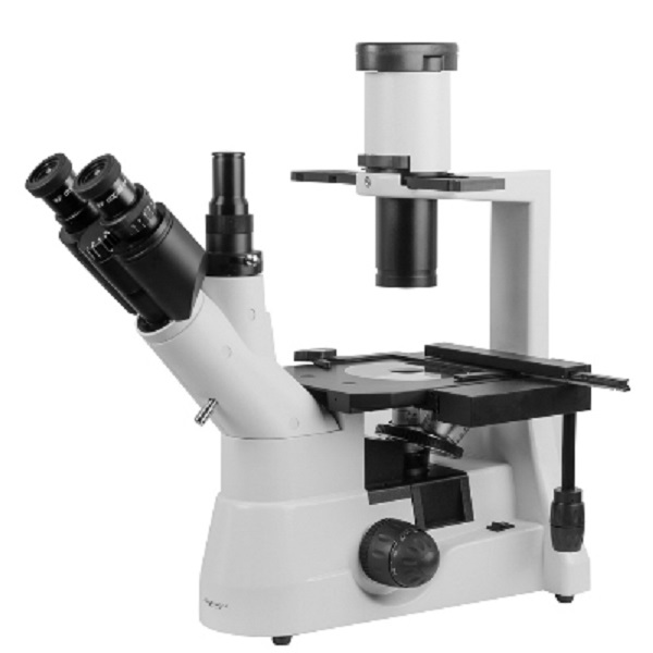
Optical microscope Micromed I
The Micromed I microscope is used for studying low-contrast tissue cell cultures, liquid sediments, etc., located in a special container. The work is carried out using the bright field method in transmitted light, as well as the phase contrast method. This model is successfully used in cell and molecular biology, pharmacology, virology and many other branches of medicine. The microscope has a design that allows you to examine not only thin samples, but also larger objects, as well as objects placed in a container with a height of 70 to 150 mm. Studies of low-contrast objects are carried out using phase-contrast lenses with magnifications of 10 RN, 20 RN. The movement of containers and utensils on the object table is done manually or using an overhead slide with coaxial control handles (included in the delivery set). For larger containers, you can increase the working surface of the object table using a special attachment (also included). The trinocular attachment will allow you to turn the model into a full-fledged digital microscope with an additional digital eyepiece.
- Microscope type: light/optical
- Attachment type: trinocular
- Magnification, times: 40-400
- Eyepieces: 10x/22
- Lenses: planachromatic: 4/0.1; 10/0.25 (phase) 40/0.6; 20/0.40 (phase)
- Interpupillary distance, mm: 50-75
- Subject table, mm: 170x240
- Backlight: halogen
- Brightness adjustment: yes
- Purpose: laboratory/medical
- Illumination location: bottom
- Research method: bright field
- Power supply: 220V/50Hz
- Dimensions, mm: 480x490x220
- Weight, kg: no more than 10.0