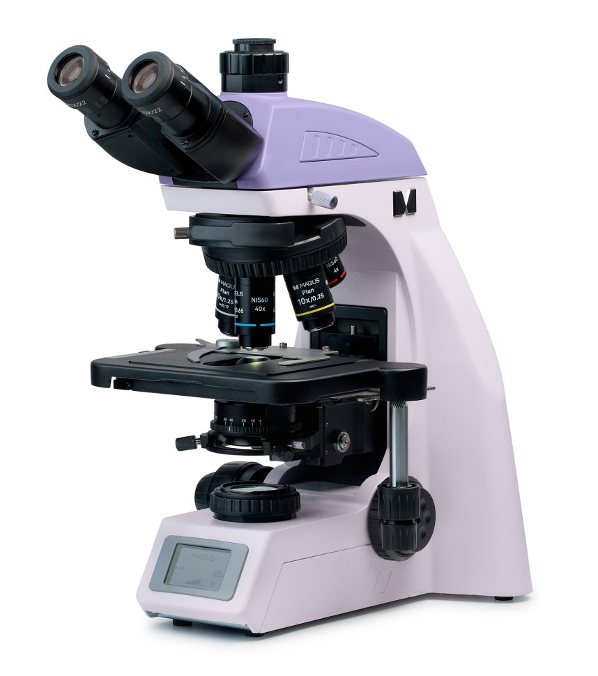
Optical microscope MAGUS Bio 260T
The MAGUS Bio 260T microscope is a routine microscope that can be used for laboratory and scientific research, or used for educational purposes. The microscope works with thin transparent and translucent specimens in a bright field. However, additional components can be installed on the device that will expand its capabilities for conducting research using dark field, phase contrast, polarized and fluorescent light methods. The model is equipped with a coded revolver, which can accommodate up to five lenses. “Smart” lighting automatically switches with the turn of the revolver and is adapted to the parameters of each lens. Thanks to this feature, there is no need to waste time adjusting the light, which is of particular importance in the educational process or in observations when you have to frequently change the magnification. The microscope has a trinocular visual head with “endless” eyepiece tubes. The height of the exit pupil is adjustable. Its position is adjusted by rotating the tubes up to 360°. A digital camera is installed in the imaging channel. The light is distributed to the camera and eyepieces in a 50/50 ratio. The microscope kit includes basic 10x/22 mm eyepieces with a diopter adjustment mechanism - no adjustment is made on the tubes. Users wearing glasses will benefit from the included rubber eyecups, which will protect the glass from abrasions and scratches. The revolver for 5 lenses is equipped with an automatic lighting control system. The fact is that when changing the magnification, the user observes strong fluctuations in the light level in the eyepiece: lenses of different magnifications transmit light in different ways. Intelligent Lighting allows you to set the light level for a specific lens once, and then when you turn the nosepiece, the light will change automatically. The microscope kit includes 4 lenses; the user can place a lens in the fifth slot in accordance with his tasks. Parfocal height – 60 mm. The connector above the turret is used to mount the analyzer; while the analyzer is not installed, the connector is closed with a plug. The object table is designed with a belt drive: the specimen moves softly and smoothly. There is no gear rack along the X axis - working with the microscope is more ergonomic and comfortable. The drug holder is fixed with two screws and can be removed when necessary. Another comfortable detail is the long table control handle. The user controls with his hands on the table, without straining his arms and body. The condenser can be adjusted to the desired position both vertically and relative to the optical axis (held on a dovetail mount). The iris diaphragm is adjusted by a ring: a marker is depicted on it, and on the condenser body there are indicators of the lens magnification. For a contrast image, the marker is set in accordance with the installed working lens. The condenser can be supplemented with dark field or phase contrast sliders: there is a special connector for this. The microscope is focused using coaxially located coarse and fine adjustment knobs. Coarse focusing is done using the handle on the left side. There are fine focusing handles on both sides of the tripod, and the right one has special convenient grooves for your fingers. Additional convenience during operation is provided by the stroke adjustment ring, which allows you to adjust the rigidity of the coarse focusing stroke in a way that is convenient for the user. All the handles and the ring are located at the bottom of the tripod: the researcher controls them by placing his hands comfortably on the table. Light in the microscope is produced by a 3-watt LED mounted in a transmitted light illuminator. The lighting temperature remains stable even with changes in brightness. Optional examination methods that can be configured on this microscope involve the use of Köhler illumination: thanks to it, the entire field of the specimen is illuminated uniformly, and artifacts such as dust or the image of the light source will not be visible in the eyepiece or digital camera. You can view the selected operating parameters and adjust them using the built-in display. It is located at the base of the microscope. The display shows the magnification of the working lens, lighting parameters, operating mode and automatic shutdown time. Additional elements will expand the functionality of the device. A set of eyepieces will increase the magnification range. For additional methods of working with drugs that cannot be seen in a bright field, it is necessary to supplement the microscope with a phase contrast device, a dark field slider, or a simple polarization device. A digital camera will help capture the results of the study in a photo or video or display the image on the screen. You can measure objects by using a calibration slide in conjunction with a scale eyepiece or a digital camera.
- Microscope type: biological
- Purpose: routine, laboratory
- Magnification, times: 40 - 1000
- Attachment type: trinocular
- Microscope attachment: with 50/50 light distribution, Gemel (Siedentopf with 360° rotation of tubes)
- Eyepiece tilt angle: 30°
- Diopter adjustment of eyepieces: ±5 diopters on both tubes
- Interpupillary distance, mm: 47 - 78
- Mounting diameter of eyepieces: 30 mm
- Eyepieces: 10x/22 mm, remote pupil
- Lenses: planachromatic, 4x/0.10, 10x/0.25, 40xs/0.65, 100xs/1.25 (oil)
- Lenses/working distance, mm: 30 (4x); 10.2 (10x); 1.5 (40xs); 0.2 (100xs)
- Parfocal height: 60 mm
- Correction for microscope tube length: infinity
- Revolving device: for 5 lenses, coded
- Stage: 230x150 mm, two-axis mechanical, without drawer
- Range of movement of the drug, mm: 78 × 54
- Condenser: Centerable and height-adjustable NA 1.25 Abbe condenser with adjustable aperture diaphragm and plug slot for darkfield and phase contrast slider
- Condenser mount: dovetail
- Diaphragm: field, aperture
- Additionally: automatic adjustment of lighting brightness when changing lenses, status display on the screen, sleep mode, eco mode
- Focusing: coaxial, coarse (30 mm, 37.7 mm/rev., with locking and rigidity adjustment mechanisms) and fine (0.002 mm, 0.2 mm/rev.)
- Backlight type: LED
- Lighting method: transmitted light
- Backlight type: LED 3W
- Backlight power: mains 220 V/50 Hz
- Research method: bright field, dark field (optional), phase contrast (optional), polarization (optional), luminescence (optional)
- Package size: 47 × 32 × 67 cm
- Package weight: 12.2 kg