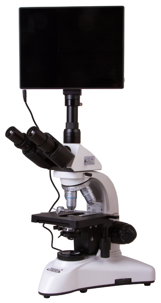
Levenhuk MED D25T LCD digital microscope
Levenhuk MED D25T LCD Digital Microscope - the presence of a 5 MP digital camera and a built-in LCD display allows you to carry out observation using the light and dark field method and demonstrate it without connecting to a PC. The planachromatic optical system allows you to magnify the image from 40 to 1000 times without loss of quality. This device is an excellent purchase for both a scientific laboratory and a medical institution. Advantage of LCD Display The presence of a display allows you to observe the material in great detail, as well as record videos and photos. In addition, by using a monitor rather than a lens as a means of observation, you will reduce eye strain, and you can also use the Levenhuk MED D25T LCD biological microscope as a visual aid in teaching, demonstrating the process in front of an audience. Observation principle The bright field method allows, without additional equipment, to examine preparations with different optical densities (both transparent and opaque) with high clarity, magnifying the image up to 1000 times without loss of quality. This method involves staining the material being studied and is therefore not suitable for studying living samples. The study of bacteria, other living microorganisms and transparent objects is possible thanks to the presence of a dark-field condenser in the kit, which can be installed in place of the standard one. When observing in this way, the material under study displays in a dark field. High-quality optics with infinite correction type Plan lenses, correcting image distortions at the edges, provide high detail of the entire object of study. As a professional category device, the microscope is equipped with an optical system, the infinite type of correction of which allows additional auxiliary elements, for example, illuminators or polarizers, to be built into the optical space between the tube and the lenses. Thus, the Levenhuk MED D25T LCD biological microscope can be optimized to solve a wide range of research problems. Thoughtful design and convenient operation The viewing part of the digital microscope can be tilted by 30° to reduce the strain on the neck and shoulders during long-term studies. Also, all optical elements of the device have a coating that protects against fungus, which is very useful for biological research. For convenient and prompt placement of the material under study under the lens, the object stage of the device is equipped with a preparation guide. You can also install additional filters in a special holder to change the contrast and improve detail. You can save the results of your work on a microSD card with a capacity of up to 32 gigabytes, which allows you to create an extensive data library. For further processing of images and videos, the package includes a CD with software that allows you to change the color parameters of the image.
- Microscope type: digital, biological
- Attachment type: trinocular
- Maximum resolution: 2048x1536 pixels
- Optics material: optical glass with anti-fungal coating
- Nozzle: 360° rotatable, with light divider
- Eyepiece tilt angle: 30°
- Magnification, times: 40-1000
- Diameter of the eyepiece tube, mm: 30 mm (binocular attachment); 23.2 mm (third vertical tube)
- Eyepieces: wide-field with diopter adjustment WF 10x/18 mm (2 pcs.)
- Lenses: planachromatic 4x, 10x, 40xs, 100xs (oil)
- Revolving device: for 4 lenses
- Interpupillary distance, mm: 55–75
- Subject table, mm: mechanical two-layer, 140x140 mm, with preparation guide
- Range of movement of the object table, mm: 75/50
- Diopter adjustment of eyepieces, D: ±5
- Condenser: Abbe N.A. 1.25 with iris diaphragm and filter holder
- Aperture: iris
- Focusing: coaxial, coarse (0.5 mm) and fine (0.002 mm), with rack and pinion mechanism
- Backlight: halogen
- Brightness adjustment: yes
- Backlight type: 6V/20W, AC 85-230V
- Light filters: blue, green, yellow
- Additionally: collector, Koehler illumination, dark field condenser (oil)
- Number of megapixels: 5
- Sensing element: 1/2.5
- Pixel size, microns: 2.2x2.2
- Sensitivity, volts per lux-second at a wavelength of 550 nm: 0.53
- Video recording capability: yes
- Frame rate: 15
- Place of use: 23.2mm microscope third eyepiece tube; using a camera mount
- White balance: auto/manual
- Exposure control: auto/manual
- Software, drivers: Android 5.1 (multilingual)
- Software capabilities: measurements, brightness, particle analysis, etc.
- Output: USB 2.0 (2 pcs.), miniHDMI, Wi-Fi, TF memory card slot
- Camera power supply: DC 12V; via AC adapter
- Additionally: LCD screen: 9.4 inches, color, touch; Built-in memory: 4 GB
- Possibility of connecting other equipment: microSD memory cards up to 32 GB; monitor/TV with HDMI connector; flash drive, computer mouse, keyboard with USB connector; 3.5 mm headphones
- Photo: *.jpg
- Video: 1080p, *.3gp
- Purpose: laboratory/medical
- Illumination location: bottom
- Research method: dark field, bright field
- Digital camera included: yes
- Case: metal
- Power supply: 100-240V