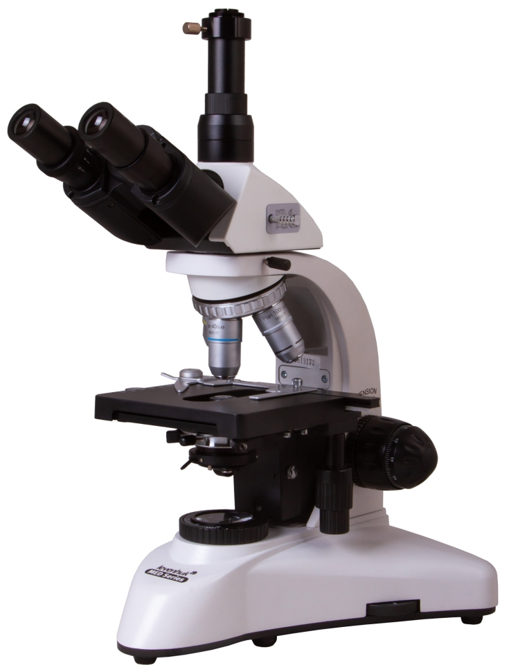
Levenhuk MED D25T digital microscope
Levenhuk MED D25T Digital Microscope is equipped with a digital camera with a resolution of 5.1 megapixels and is designed for observation using the light and dark field method in a magnification range from 40 to 1000 times. You can use the device as a visual aid by connecting the microscope to an external monitor using a USB cable. This device is an excellent purchase for both a scientific laboratory and a medical institution. Observation principle The bright field method allows, without additional equipment, to examine preparations with different optical densities (both transparent and opaque) with high clarity, magnifying the image up to 1000 times without loss of quality. This method involves staining the material being studied and is therefore not suitable for studying living samples. The study of bacteria, other living microorganisms and transparent objects is possible thanks to the presence of a dark-field condenser in the kit, which can be installed in place of the standard one. When observing in this way, the material under study displays in a dark field. What are the advantages of a digital model? By connecting the camera to a PC using a USB cable, you can broadcast the research process online to an external monitor. Thus, the Levenhuk MED D25T digital microscope can become a visual aid in the educational process, and also allows you to involve a group of people in research. In addition, using a wide screen rather than a lens as a means of observation, you will reduce eye strain and be able to see the smallest details. High-quality optics with infinite correction type As a professional category device, a digital microscope is equipped with an optical system, the infinite type of correction of which allows additional auxiliary elements, for example, illuminators or polarizers, to be built into the optical space between the tube and the lenses. Thus, the microscope can be optimized to solve a wide range of research problems. Thoughtful design and convenient operation The viewing part of the Levenhuk MED D25T biological microscope is tilted by 30° to reduce the load on the neck and shoulders during long-term studies. Also, all optical elements of the device have a coating that protects against fungus, which is very useful for biological research. For convenient and prompt placement of the material under study under the lens, the object stage of the device is equipped with a preparation guide. You can also install additional filters in a special holder to change the contrast and improve detail. You can save your work on a removable microSD card with a capacity of up to 32 GB, which allows you to create an extensive data library. For further processing of images and videos, the package includes a CD with software that allows you to change the color parameters of the image.
- Microscope type: digital, biological
- Attachment type: trinocular
- Maximum resolution: 2048x1536 pixels
- Optics material: optical glass with anti-fungal coating
- Nozzle: 360° rotatable, with light divider
- Eyepiece tilt angle: 30°
- Magnification, times: 40-1000
- Diameter of the eyepiece tube, mm: 30 mm (binocular attachment); 23.2 mm (third vertical tube)
- Eyepieces: wide-field with diopter adjustment WF 10x/18 mm (2 pcs.)
- Lenses: planachromatic 4x, 10x, 40xs, 100xs (oil)
- Revolving device: for 4 lenses
- Interpupillary distance, mm: 55–75
- Subject table, mm: mechanical two-layer, 140x140 mm, with preparation guide
- Range of movement of the object table, mm: 75/50
- Diopter adjustment of eyepieces, D: ±5
- Condenser: Abbe N.A. 1.25 with iris diaphragm and filter holder
- Aperture: iris
- Focusing: coaxial, coarse (0.5 mm) and fine (0.002 mm), with rack and pinion mechanism
- Backlight: halogen
- Brightness adjustment: yes
- Backlight type: 6V/20W, AC 85-230V
- Light filters: blue, green, yellow
- Additionally: collector, Koehler illumination, dark field condenser (oil)
- Number of megapixels: 5.1
- Sensing element: 1/2.5
- Pixel size, microns: 2.2x2.2
- Sensitivity, volts per lux-second at a wavelength of 550 nm: 0.53
- Video recording capability: yes
- Frame rate: 7@2592x1944 27@1280x960 90@640x480
- Dynamic range, dB: 66
- Use location: 23.2mm microscope third eyepiece tube
- Spectral range, nm: 380–650
- White balance: auto/manual
- Exposure control: 0.294–2000 ms
- Software, drivers: Levenhuk
- Software features: image size, brightness, exposure time
- Output: USB 2.0, 480 Mbps
- System requirements: Windows XP/Vista/7/8/10 (32 and 64 bit), Mac OS 10.12, Linux, 2.8 GHz Intel Core 2 or higher, at least 2 GB RAM, USB 2.0, 17 monitor
- Camera power supply: via USB cable
- Illumination location: bottom
- Research method: dark field, bright field
- Digital camera included: yes
- Case: metal
- Power supply: 100-240V