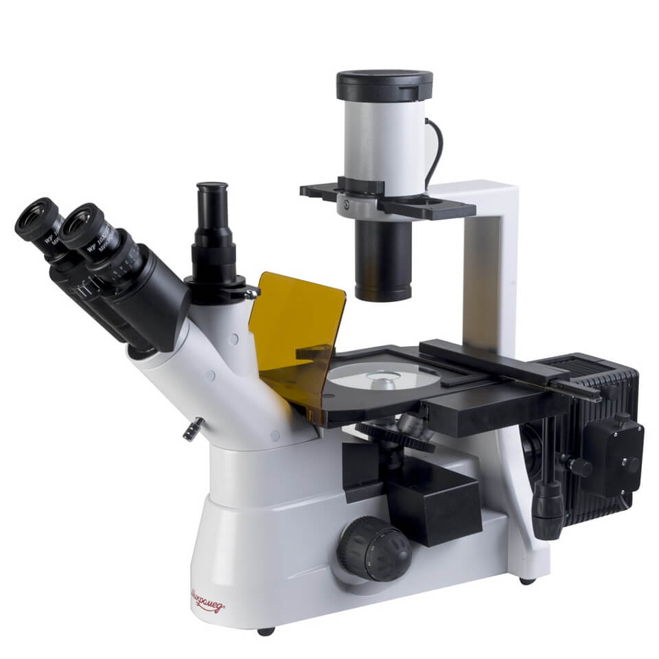
Microscope Micromed AND LUM (inverted, luminescent)
Microscope Micromed AND LUM (inverted, luminescent). Trinocular inverted fluorescent microscope is designed for studying low-contrast tissue cell cultures and liquid sediments in special containers. Objects are studied in transmitted light using the bright field and phase contrast methods, as well as in the light of visible luminescence. The microscope can be used in the field of medicine, cellular and molecular biology, biotechnology, pharmacology, toxicology, virology, hydrobiology, agriculture, ecology. The inverted design of the microscope, in which the object is illuminated from above and observed from below, allows you to examine large objects or objects located in special dishes (Petri dishes, Terrasaki plates, flasks), as well as view the nutrient medium under the monolayer. The lighting system of the microscope is designed to work with laboratory glassware with a height of up to 70 mm; it is possible to install glassware with a height of up to 150 mm. The lenses of the plan achromatic correction microscope have an increased working distance, which allows you to view objects in laboratory dishes with a bottom thickness of up to 1.5 mm, as well as view the nutrient medium above the monolayer. The eyepieces have a field of view of 22 mm, diopter vision correction and “remote pupil”, which allows you to work equally comfortably both with and without glasses. Studies of low-contrast objects are carried out using phase-contrast lenses with magnifications of 10 RN, 20 RN. The luminescence module contains 2 standard luminescence cubes (blue and green). The third open position allows the user to quickly switch from the fluorescence method to the bright field method. The glassware is moved on the object table manually or by an overhead slide with coaxial handles. On the microscope, you can photograph images of objects using a f/c-based visualization kit (not included) and display the image in real time on a PC screen using a video eyepiece (not included). The video eyepiece is installed in the third vertical output of the visual attachment. Features: Modern ergonomic design. The optical design of the microscope is designed for infinity. The eyepieces have a field of view of 22 mm, diopter vision correction and “remote pupil”. Base with built-in power supply and dimmable illuminator. High-precision assembly, microscope alignment and parfocal objectives. Two-coordinate stage with coaxial handles. Coaxial coarse and fine focusing mechanism. Coarse focusing locking mechanism for quick adjustment of the microscope when changing specimens. Adjusting the rigidity of the coarse focusing stroke. Manba: https://www.nv-lab.ru/catalog_info.php?ID=5403
- Magnification, times: 40 - 400
- Spectral range of luminescence excitation, nm: 515-700
- Spectral range of the studied luminescence, nm: 410-550
- Visual Head: Trinocular
- Nozzle increase: 1
- Diopter adjustment (on the right tube), D: ±5
- Mounting diameter of eyepieces, mm: 30
- Tilt angle of the visual nozzle, degrees: 45
- Adjustable interpupillary distance, within, mm: 50-75
- Eyepieces: wide-field with 10/22 eye relief
- Revolving device: for 5 lenses
- Lens correction type: long-focus planchromats, for working in visible luminescence light,
- designed for tube length "infinity"
- Lenses: 4x/0.1
- 10x/0.25 phase
- 20x/0.40 phase
- 40x/0.6
- Subject table, mm: 170x240
- Range of movement of the drug, mm: 80x120
- Condenser, NA: 0.3
- Source of transmitted light - halogen lamp, W/W: 12/30
- Fluorescent light source - mercury lamp, W: 100
- Power source - AC mains, V/Hz: 220±22/50
- Overall dimensions, mm: 700 x 320 x 500
- Weight, no more, kg: 10