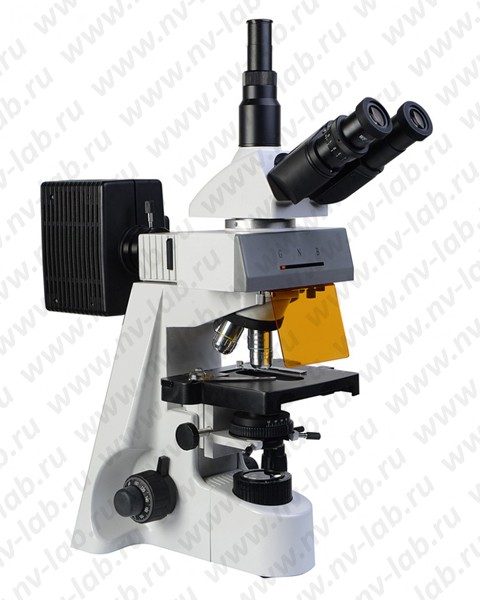
Microscope Mikromed-3 LUM (trinocular, luminescent)
Trinocular luminescent microscope Mikromed 3 LUM is intended for observing images of objects in the light of visible luminescence, as well as in transmitted light in a bright field. Areas of application: • Medicine (morphological, cytological, molecular genetic, immunological, microbiological research methods); • Biology • Veterinary • Food and pharmaceutical industry • Forensic science • In the field of sanitary and epidemiological surveillance • In the field of environmental protection • Geology In addition to the possibility of studying objects in transmitted light (classical Keller illumination), using fluorescent techniques on a Micromed 3 microscope LUM conducts the following types of studies: immunochemical, immunological, immunomorphological, immunogenetic. In the process of studying drugs - blood smears, bone brain, tissue sections is carried out: identification of hidden infections, such as chlamydia, ureaplasmosis, mycaplasmosis, herpes and others; and also: • identification of differentiation antigens of T and B lymphocytes, atypical cells blood; • express diagnostics of bacterial, viral, protozoal, etc. infections; • determination of antinuclear factor, etc.; • immunochemical diagnosis of leukemia; • chromosome analysis. The microscope includes: a tripod with source lights light, revolving lens mounting device, object stage and condenser; trinocular attachment; mercury lamp power supply; kit lenses; eyepieces and filters; set of spare parts; kit accessories. The operating principle of the microscope is based on the use of the phenomenon luminescence of observed objects arising under the influence of light certain spectral composition. Illumination of objects for excitation Luminescence is produced from above through an opaque illuminator and a microscope lens. The light source is a mercury lamp. Light needed for stimulation luminescence, is isolated from the general radiation of a mercury lamp using light filters, conventionally called excitation light filters. Light is directed into the lens by a beam splitter with interference coating, predominantly reflecting the excitation light and transmitting the luminescent light of the object. Excitation filter, The beam splitter and cutting filter are combined into a fluorescent filter block Advantages: - Ergonomic tripod design with coaxial coarse and fine focusing knobs - Improved image quality and comfort in observing the object being examined due to the use of high-quality optics, providing flat and microscope field of view increased to 22 mm. - Possibility of using visualization systems - Low price for a microscope due to economical basic configuration Manba: https://www.nv-lab.ru/catalog_info.php?ID=1118
- Microscope magnification, times:
- Fluorescent block:
- Light source:
- Power source - AC mains
- smallest - 40x
- highest - 1000x
- - for various methods of fluorescent analysis in
- microscopy
- - spectral range of luminescence excitation, nm:
- 410-550
- - spectral range of the studied luminescence,
- nm: 515-700
- - filter system in the main block: 2 exciting
- filters, dichroic mirror, 2 cut-off filters
- - filter blocks: B (blue), G (green), N (passive
- light)
- - protective screen
- - mercury lamp 100W
- - power supply 220 V 50/60 Hz
- Visual attachment
- Angle of inclination of the visual nozzle, degrees:
- Increased visual
- nozzles
- Revolving device:
- Trinocular
- 45
- 1
- Reverse revolver for four lenses
- Lenses: E Plan (length of the tube is designed for
- "infinity")
- 4x/0.10 ∞/0.17
- 10x/0.25 ∞/0.17
- 40x/0.65 ∞/0.17 (spring loaded)
- 100x/1.25 MI ∞/0.17 (spring loaded)
- Adjusting the interpupillary distance
- within, mm:
- Diopter compensation:
- 55-75
- +/- 5 dioptres
- Wide-field eyepieces, magnification/field
- WF 10x DIN 22
- Centerable Abbe condenser with iris diaphragm and
- filters
- Focus:
- Micrometric focusing mechanism vernier:
- Ability to work with both hands:
- Height adjustable, with iris diaphragm, nA 1.25, s
- filter holder
- - coaxial coarse and fine focusing screws
- - Built-in focus locking mechanism for protection
- drug and quick adjustment when changing it
- - smoothness adjustment mechanism
- - rack and pinion focusing mechanism
- - 0.002 mm
- yes
- Subject table
- Rectangular size 180x150mm
- drug handle on the right
- Range of movement of the drug, mm:
- Tripod:
- 75x50
- metal, with rubber feet, painted
- fireproof enamel
- Transmitted light source:
- Halogen lamp 6V, 20W
- Power supply:
- fuses
- Power supply built into the tripod
- 220 V 50 Hz
- 1 A
- Operating temperature:
- Humidity:
- 18 - 35°C
- less than 80%
- Weight of the microscope together with the block, no more, kg
- Power source weight, kg:
- Overall dimensions, mm:
- 9.8
- 1.5
- without packaging: 220x270x510
- in packaging: 370x460x520