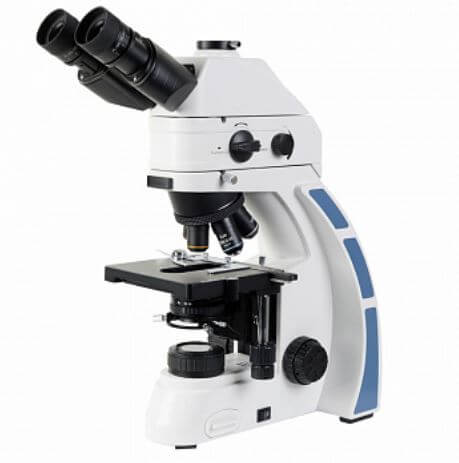
Microscope Micromed 3 Alpha luminescent, 21779
Microscope Micromed 3 Alpha luminescent, 21779 The easy-to-use LED fluorescence microscope allows for fast research. Benefits: quick switching from fluorescence to bright field; LEDs with stable color temperature and high efficiency; ease of use; Possibility of use for simple laboratory research; modular design allows you to expand the microscope for conducting research in the dark field, using the phase contrast method, in polarized light. With fluorescent microscopy, there is significantly greater sharpness and contrast of the microscopic picture, which increases the comfort of the study. It is much easier for the researcher's eye to detect fluorescent bright yellow-green mycobacteria on a dark, almost black background than to detect pink mycobacteria on a blue background using Ziehl-Neelsen staining. This makes the fluorescent microscopy method especially valuable when studying oligobacillary material. The sensitivity of fluorescent microscopy, especially in combination with the method of enrichment of diagnostic material (sediment microscopy), approaches the sensitivity of the culture method. These advantages make it possible to use fluorescence microscopy in laboratories that perform a large number of studies daily (about 30 or more) in order to speed up the microscopy procedure. The luminescent attachment of the Micromed 3 Alpha microscope is designed specifically for the study of Koch's bacillus (Auramine O staining) and does not require the selection of light filters, which minimizes the possibility of obtaining false positive results. In all doubtful cases of microscopic picture, microscopy of a smear stained using the Ziehl-Neelsen method should be used for control. Microhoney 3 Alpha easily switches to work in transmitted light and does not require additional settings. Thus, it replaces 2 microscopes in the laboratory: for working in luminescence light (staining a smear with Auramine O) and for working in transmitted light (for examining a smear stained using the Ziehl-Neelsen method). Manba: https://www.nv-lab.ru/catalog_info.php?ID=8673
- Parameter: Value
- Microscope magnification, x: 40 – 1000
- Excitation (EX): 450/50
- Dichroism (DI): 480LP
- Emission (EM): 485LP
- Visual attachment: design – Gemel;
- trinocular;
- tilt angle 30°;
- 360° rotatable;
- 80/20 light split
- Adjustable interpupillary distance, within, mm: 50 – 75
- Nozzle increase: 1
- Eyepieces: wide-field with 10/22 eye relief
- Revolving device: for 5 lenses
- Type of lens correction: planchromatic, for working in visible luminescence light, designed for tube length "infinity"
- Lenses, magnification / aperture: 4× / 0.1
- Stage size, mm: 150 × 140
- Range of movement of the drug, mm: 76 × 50
- Condenser device: Abbe condenser with a hinged frame, with slots for installing additional devices (polarization, dark field, phase contrast), with aperture markings for lenses
- Transmitted light source: 3W LED
- Fluorescent light source: LED – blue LED (455 nm; 3 W / 6 V)
- Power source – AC mains, V/Hz: 220±22/50
- Overall dimensions, mm: 400 × 200 × 500
- Weight, kg, no more: 10