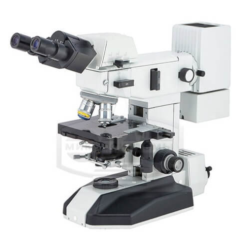
Microscope Mikmed-2 version 11 luminescent
Microscope Mikmed-2 version 11 luminescent. Microscope for clinical laboratory diagnostics MIKMED-2 (fluorescent) is intended for studying objects using fluorescent contrast methods (when illuminated from above through the lens) and colored preparations in transmitted light (classical Keller illumination). A set of spectral units with beam splitting plates, excitation and cut-off filters is designed for the study of objects labeled with Fluorescein isothiocyanate (FITC), as well as treated with dyes, such as: DAPI Hoechst, Auramine, Acridine orange, Rhodamine, Propidium iodide, etc. Research is carried out in transmitted light: cytological, morphological, molecular genetic, immunological, microbiological Manba: https://www.nv-lab.ru/catalog_info.php?ID=4206
- Apparent magnification of the microscope, x: 100 - 1500
- Spectral range of luminescence excitation, nm: 450 - 550, (360* - 550)
- Spectral range of the studied luminescence, nm: 520* - 700, (400* - 700)
- Beam splitter guides: Green-2, Red, Blue*, Green*
- Visual attachment: trinocular (beam separation, %: binocular/adapter 100/0 or 0/100);
- Nozzle magnification, x: 1
- Tilt angle of eyepiece tubes, degrees: 20
- Adjustable interpupillary distance, mm: 55-75
- Eyepieces: apparent magnification, magnification/field, mm: Wide-field
- 10x/18 or 10x/20
- 15x/12 or 15x/15
- Revolving lens mount: Four-socket, rotation in any direction
- Lens correction type: Microfluars, stigmachromats
- Objectives: (magnification), multiple/numerical aperture: Microfluars:
- Stigmachromats:: 20/0.45L ∞/0.17 OSKH-20L-0
- 40/0.65L ∞/0.17 OSKH-40L-0
- for objects without cover glass: 40/0.65L ∞/0 OSKH-40LB-0
- Two-coordinate stage: coaxial handles, control on the right
- Range of movement of the drug, mm: 76x26
- Focusing mechanism for moving the table: coaxial handles for coarse and fine focusing -
- on both sides of the tripod, coarse tightness adjustment handle
- focusing - left, height movement limitation
- Condenser: centered, focusable, numerical aperture A=0.9,
- iris diaphragm
- Lighting: transmitted light built-in classic according to Keller
- with centered and focused iris field diaphragm
- Light sources:
- Transmitted light
- Incident light: white LED 5 W
- 100 W mercury lamp
- Power source: AC mains, 220 V 50 Hz,
- The LED power supply is built into the tripod,
- Provides smooth brightness adjustment
- Overall dimensions, mm:
- Microscope
- Mercury lamp power source: 220x530x440
- 130x220x80
- Weight, kg:
- Microscope
- Mercury lamp power supply: 17
- 1.4