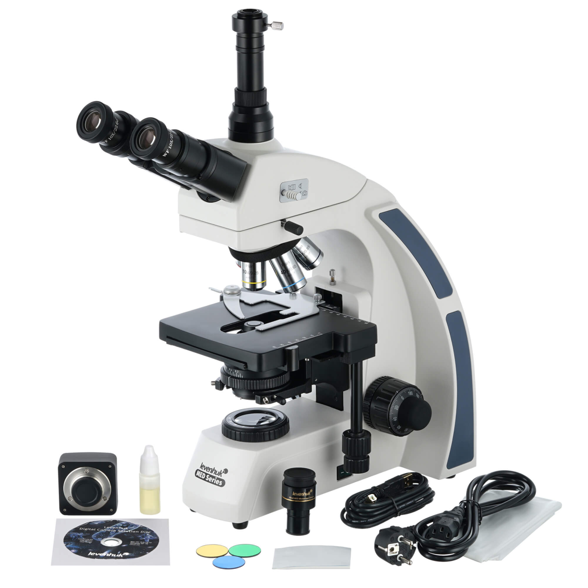
Levenhuk MED D40T digital trinocular microscope
Levenhuk MED D40T digital trinocular microscope Article: 74007 Levenhuk MED D40T is a microscope with a 16 MP digital camera. It can be used not only for visual observations, but also for creating digital archives of research. Professional-grade optics allows you to study complex and extended microstructures. Allows you to conduct research in microbiology, biochemistry, medicine, veterinary medicine and other fields of science. "Infinity" high-end optics Levenhuk MED 40 series microscopes are equipped with an optical system that is adjusted to infinity, which is used in professional and high-end microscopes. This system includes Infinity PlanAchromat lenses and produces clear, high-contrast images with a high degree of flatness. One of the most important features of the infinity optical system is the ability to install in the optical path of the microscope between the objectives and the eyepiece. tube any additional components. These include polarizers and epi-fluorescent illuminators. As a result, the modular design principle and ease of operation make Levenhuk MED 40 optimal microscopes for using different types of microscopy and working in hematology, histology, microbiology and other laboratories. Rotating trinocular head with light splitter The microscope uses a trinocular attachment with a light splitter. It allows you to use the light flux most efficiently, dividing it between the binocular visual unit and a separate eyepiece tube designed for mounting the camera. The head can be rotated 360°, making group observations more convenient. And a slight tilt of the visual unit by 30° reduces the load on the neck muscles - long-term observations cause less fatigue. Visual observations without distortion The microscope uses plan achromatic optics: five objectives provide different magnifications and allow observations using the “dry” method. In addition to the 100x objective, it can also be used for oil immersion. An immersion lens will allow for maximum multiplicity, it is better to examine the details of microslides, greatly enhancing their brightness and depth of field. A bottle of immersion oil is included in the kit. To protect the optics, the three lenses with the highest magnification are equipped with spring-loaded frames. The eyepieces are wide-field and have diopter adjustment. They zoom in 10 times. All optics are made of glass with anti-fungal coating. Digital capabilities The package includes a 16-megapixel camera that can be connected to a computer. It allows you to take photos, record videos, and broadcast. The image from the camera is displayed on the connected screen in real time. Thanks to this, during research there is no need to constantly look into the eyepieces, straining your eyesight and neck muscles, so long work in the laboratory becomes lighter and more convenient. Using a special program, you can adjust the size, brightness and contrast of the image, change the exposure time, calibrate the camera with lenses, measure samples or individual structures (several units of measurement are available). Convenient work with micropreparations The stage can be moved in two directions. It is equipped with a preparation guide that helps to accurately place samples under the lens. Next to the table are the coarse and fine focusing knobs. This method of adjusting sharpness used exclusively in the highest class microscopes. Under the table there is a bright LED backlight and an Abbe condenser with an iris diaphragm, which allows you to select the appropriate aperture to accurately direct the light beam to the observation area. The included filters help highlight details and increase image contrast. The brightness of the backlight can be adjusted; AC power is used for power supply. You can use lighting adjusted according to the Köhler method. Features: 16 megapixel digital camera included. Trinocular microscope, attachment with light splitter. “Infinite” plan achromatic lenses. Magnification range – from 40 to 1000 times. Bright LED backlight. Possibility of customizing lighting using the Köhler method. Powered by AC mains, backlight brightness adjustable. Scope of delivery: 16 MP digital camera (Adapter for camera, USB cable, Disk with software and drivers). Microscope stand with base. Trinocular 360° rotatable head. Plan achromatic lenses, infinity corrected: 4×, 10×, 40×s, 60×s, 100×s (oil) with anti-fungal coating. Wide-field eyepieces: WF10×/22 mm with anti-fungal coating (2 pcs.). Abbe condenser N.A. 1.25. Iris diaphragm. Filters: blue, green, yellow. Bottle with immersion oil. Fuse (2 pcs.). Power cord for microscope. Dust cover. Operating instructions. Warranty card. Manba: https://www.nv-lab.ru/catalog_info.php?ID=8634
- Parameter: Value
- Microscope type: digital, light/optical, biological
- Attachment type: trinocular
- Optics material: optical glass with anti-fungal coating
- Nozzle: 360° rotatable, with light divider
- Eyepiece tilt angle: 30°
- Magnification, times: 40 – 1000
- Diameter of the eyepiece tube, mm: Ø30 (binocular attachment);
- Ø23.2 (third vertical tube)
- Eyepieces (paired): WF10×/22 mm (wide-field with diopter correction)
- Lenses: planachromatic, infinity corrected: 4×, 10×, 40×s, 60×s, 100×s (oil)
- Revolving device: for 5 lenses
- Interpupillary distance, mm: 48 – 75
- Subject table, mm: 180×150, mechanical two-layer, with preparation guide
- Range of movement of the object table, mm: 75 / 50
- Diopter adjustment of eyepieces, D: ±5
- Condenser: Abbe N.A. 1.25
- Aperture: iris
- Focusing: coaxial, coarse (0.5 mm) and fine (0.002 mm), with rack and pinion mechanism
- Case: metal
- Backlight / Backlight lamp type: LED / LED 5 W
- Brightness adjustment: yes
- Power supply: 85-240V/50Hz
- Light filters: blue, green, yellow
- Additionally: collector, Köhler lighting
- Number of megapixels: 16
- Sensing element: 1/2.33”
- Pixel size, microns: 1.335 × 1.335
- Maximum resolution: 4632 × 3488
- Frame rate: 2 frames at a resolution of 4632 × 3488;
- 8 frames at a resolution of 2320 × 1740;
- 11 frames at 1536 × 1160 resolution
- Photo: *.jpg, *.bmp, *.png, *.tif, etc.
- Video recording capability: yes
- Video: *.wmv, *.avi, *.h264 (Windows 8 and above);
- *.h265 (Windows 10 and above)
- Place of use: microscope eyepiece tube, instead of eyepiece
- Spectral range, nm: 380 – 650
- White balance: auto / manual
- Exposure control: 0.2 – 2000 ms
- Exposure control: auto/manual
- Software, drivers: LevenhukLite
- Software features: image size, brightness, exposure time
- Output: USB 2.0
- System requirements: Windows 8/10/11 (32 and 64 bit), macOS X, Linux, up to 2.8 GHz Intel Core 2 and higher, 2 GB RAM, USB 2.0 port, CD-ROM
- Camera power supply: via USB cable
- Optional magnification options: The magnification range can be expanded by using optional. accessories (eyepieces, Barlow lenses, objectives)
- User level: for experienced, for professionals
- Assembly and setup difficulty level: difficult
- Purpose: laboratory / medical
- Illumination location: bottom
- Research method: bright field
- Digital camera included: yes
- Cover/case/bag included: dustproof cover
- Package size (L × W × H), mm: 500 × 440 × 280
- Package weight, kg: 10.34