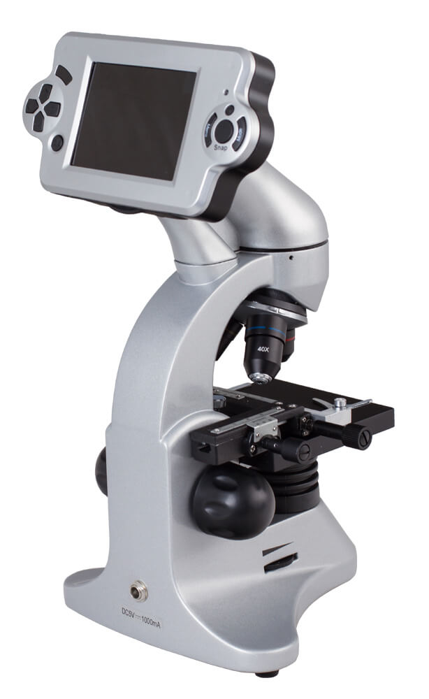
Levenhuk D70L digital monocular microscope
Levenhuk D70L digital monocular microscope Article: 14899 The Levenhuk D70L digital biological microscope is well suited for teaching schoolchildren and students; it is convenient to use in the educational process - the image of a microscopic specimen is displayed on an LCD monitor, where it can be seen by everyone present at the same time during group work. High quality optics provide a bright and contrasting image; this microscope model allows you to observe and save images of microscopic specimens and opaque objects using the bright field method. Microscope allows you to quickly and efficiently carry out educational work in biology and other sciences that require the study of microscopic objects. High image quality All optical elements of the microscope are made of high-quality glass and coated with a multilayer antireflective coating. This provides the brightest and most contrasting image without distortion across the entire field of view. The microscope allows you to obtain optical magnification in the range of 40 to 400 times, and the digital zoom allows you to obtain magnification up to 1600 times. LCD monitor The image of microspecimens is recorded by a 2 megapixel camera and displayed on an LCD monitor with 3.6" (9.1 cm) diagonal in real time. Turret for lenses The microscope lenses are mounted on a revolving head, which makes it possible quickly change the magnification used while working. In order to move to another magnification, it is enough to rotate the turret around its axis - this takes only a few seconds. Built-in backlight The microscope is equipped with two built-in illuminators, which allow you to provide sufficient illumination of microspecimens even when working in a dark room. LEDs are used as light sources - unlike incandescent lamps, they have a long service life (tens of thousands of hours) and do not burn out from mechanical stress. The brightness of the LEDs is adjustable, which allows you to select optimal lighting conditions for each microslide. Ability to observe opaque objects The microscope has an upper illuminator - this makes it possible to examine opaque objects in reflected light. This will allow us to examine the microstructure of such familiar and everyday things as a sheet of paper, a coin, etc. Observations are carried out using the bright field method. Main features: Coated glass optics. Ability to save images to a memory card. Magnification from 40 to 1600 times. Camera with a resolution of 2 megapixels. LCD screen with a diagonal of 3.6" (9.1 cm) and a CMOS matrix. Built-in LED lights. Allows you to examine opaque objects. Turret head for lenses. Equipment: Microscope. Lenses: 4×, 10× and 40×. Eyepiece: WF10×. Adapter for eyepiece. Removable LCD display with memory card slot. SD memory card 8 GB. USB cable. Subject stage with clamps and coordinate movement. Disc with apertures and five color filters. Built-in bottom and top LED lights. Power cord 220 V. Guide “Interesting microscope. We study the microworld." Tweezers. Incubator for Artemia. A microtome is a tool for obtaining thin sections from samples and preparations examined under a microscope, allowing you to obtain sections with a thickness of fractions millimeter. A bottle of yeast. A bottle with resin for making drugs. Bottle of sea salt. A bottle with Artemia is a sea crustacean that is common in the seas and oceans of the whole world, used as food for aquarium fish, as well as in fish factories. 5 ready-made samples and 5 clean slides. Dust cover. Plastic case. Operating instructions Warranty card. Manba: https://www.nv-lab.ru/catalog_info.php?ID=8635
- Parameter: Value
- Microscope type: digital, light/optical, biological
- Attachment Type: Digital Display/PC Monitor
- Optics material: optical glass
- Nozzle: fixed (non-rotating)
- Magnification, times: 40 – 1600
- Eyepiece tube diameter, mm: Ø23.2
- Eyepieces: WF10×
- Lenses: 4×, 10×, 40×
- Revolving device: for 3 lenses
- Interpupillary distance, mm: 54 – 75
- Subject table, mm: 90×95 with preparation guide
- Range of movement of the object stage, mm: 0-10 vertically
- Condenser: N.A. 0.65
- Focus: Coarse 13mm
- Case: metal
- Backlight: LED
- Brightness adjustment: yes
- Power supply: 220V/50Hz or 2 AA batteries (not included)
- Number of megapixels: 2
- Photo: *.jpg, *.bmp, *.png, *.tif, etc.
- Video recording capability: yes
- Software, drivers: pre-installed Android system
- Ability to connect other equipment: Windows OS and macOS computers for copying files
- Output: USB 2.0, AV
- User level: for beginners
- Assembly and setup difficulty level: easy
- Purpose: school/educational
- Backlight location: combined (top and bottom)
- Research method: bright field
- Digital camera included: yes
- Experiment kit included: yes
- Case/case/bag included: case, dust cover
- Package size (L × W × H), mm: 400 × 260 × 170
- Package weight, kg: 3.52